")
Back to Journals » Clinical Interventions in Aging » Volume 14
Effect of 12-week home-based cognitive training on cognitive function and brain metabolism in patients with amnestic mild cognitive impairment
Authors Park J , Kim SE, Kim EJ , Lee BI, Jeong JH, Na HR, Choi SH , Kang DY , Park KW
Received 3 January 2019
Accepted for publication 22 May 2019
Published 28 June 2019 Volume 2019:14 Pages 1167—1175
DOI https://doi.org/10.2147/CIA.S200269
Checked for plagiarism Yes
Review by Single anonymous peer review
Peer reviewer comments 4
Editor who approved publication: Dr Richard Walker
Jinse Park,1 Si-Eun Kim,1 Eun-Joo Kim,2 Byung In Lee,1 Jee Hyang Jeong,3 Hae Ri Na,4 Seong Hye Choi,5 Do-Young Kang,6 Kyung Won Park7
1Department of Neurology, Haueundae Paik Hospital, Inje University, Busan, South Korea; 2Department of Neurology, Pusan National University Hospital, Pusan National University School of Medicine and Medical Research Institute, Busan, South Korea; 3Department of Neurology, Ewha Womans University Mokdong Hospital, Ewha Womans University School of Medicine, Seoul, South Korea; 4Department of Neurology, Bobath Memorial Hospital, Seongnam, South Korea; 5Department of Neurology, Inha University School of Medicine, Incheon, South Korea; 6Department of Nuclear Medicine, College of Medicine, Dong-A University, Busan, South Korea; 7Department of Neurology, Cognitive Disorders and Dementia Center, Dong-A University College of Medicine and Institute of Convergence Bio-Health, Busan, South Korea
Purpose: We assessed the effect of home-based cognitive intervention (HCI) on cognitive function along with brain metabolism by 18F-FDG PET in patients with amnestic MCI (aMCI).
Patients and methods: Fifty-seven patients with aMCI from three hospitals were randomized (30 HCI, 27 control). For 12 weeks, subjects received HCI. Thirty-two subjects (15 HCI, 17 control) underwent brain 18-F-FDG-PET imaging at baseline and at 12 and 24 weeks.
Results: The HCI group showed significant improvement in the scores of the Controlled Oral Word Association Test (COWAT) 12 and at 24 weeks. Significant brain metabolic changes by 18F-FDG PET were not observed.
Conclusion: The current study suggests that HCI was effective in improving general cognition along with frontal executive function in patients with aMCI.
Keywords: mild cognitive impairment, cognitive training, brain PET
Introduction
With an increasingly aging society, the percentage of the population with cognitive decline is rising rapidly. Mild cognitive impairment (MCI) is a heterogenic condition that has a high risk of conversion to Alzheimer’s dementia (AD);1 therefore, MCI is a target for prevention of development of AD.2 Pharmacological therapy such as acetylcholinesterase inhibitors can only delay disease progression in AD up to 2 years and new drug trials have failed.3 Clinical trials for preventing conversion of MCI to AD have yielded inconsistent results, so a multifactorial approach to prevent dementia in patients with MCI has been attempted.4,5
Non-pharmacological treatments including cognitive training and stimulation have emerged and received much attention.6,7 Many studies show the effectiveness of cognitive training in AD. However, there is a lack of high-quality randomized controlled trials.8,9 Amnestic MCI (aMCI) is more likely than non-amnestic MCI to progress to AD.10 Previous reports have demonstrated that cognitive training has a more beneficial effect in adults with normal cognition or MCI state than in those with AD.8,11
In Korea, a Korean cognitive training program consisting of a paper-based quiz was developed and demonstrated a positive effect on cognition in aMCI.12 However, its clinical application is limited by cost and space factors. Home-based cognitive training can overcome these limitations, increasing adherence. So far, studies have not compared the effectiveness of home-based individual versus hospital-based group training. We investigated the effectiveness of home-based cognitive training in aMCI patients.
Several studies have sought to investigate the mechanism of cognitive training in patients with dementia. Jung et al found that serum BDNF level was decreased after 12 weeks' home-based cognitive training. Another common approach to investigating the mechanism of intervention is by using functional imaging. There are few published studies investigating the effect of cognitive training on brain metabolism in MCI. 18F-FDG PET can assess brain metabolism, a well-established biomarker in cognitive dysfunction.13 Foster et al investigated the change in glucose metabolism in the frontotemporal area and corpus callosum by FDG PET study after cognitive training.14 Another study demonstrated that cognitive stimulation induced a change in Brodmann area in 18F-FDG PET in MCI patients.15 We evaluated the change in brain metabolism by 18F-FDG PET as well as cognition after cognitive training.
Materials and methods
Study design
This was a prospective, multicenter, randomized, open-labelled, placebo-controlled clinical trial. Patients were recruited from three different centers including Dong-A University, Busan National University Hospital, and Haeundae Paik Hospital. These are the biggest hospitals, and widely spread, in Busan city in Korea. n terms of patients, there is no difference in race, population and basic demographic features among the three centers. The study was approved by the Institutional Review Board in each hospital and conducted in accordance with the Declaration of Helsinki.
Fifty-seven patients with aMCI diagnosed by NIA-AA criteria were enrolled and randomly assigned in a 1:1 ratio to the home-based (31) and control (26) groups using a random-number table (permuted block randomization). Allocation concealment was delivered to each interviewer in a sealed enveloped from the CRO company. Randomization was requested by the interviewer responsible for recruitment and interview. The study was open-label, so we did not conceal the block size until analysis of primary outcome. When interventions had been completed, clinical data were delivered to the study coordinator in Dong-A university.
We used an established cognitive training program in Korea.12 The training period was 30 mins, daily, for 12 weeks.
Subjects
Inclusion criteria were as follows: 1) clinical diagnosis of aMCI;16 2) decreased Seoul verbal learning test, < standard deviation; 3) normal activity (≤7 in Seoul Instrumental Activities of Daily Living); 4) no clinical diagnosis of dementia; 5) score of < on the Hachinski ischemic scale; 6) literate; 7) age 50–80 years; 8) no organic brain lesions.
Exclusion criteria were as follows: 1) participation in other clinical trials or receiving medication for a clinical trial within 4 weeks; 2) serious medical problems that could interfere with the study; 3) an accompanying disease that might affect the neuropsychological test; 4) other neurodegenerative and psychiatric diseases; 5) a history of alcohol dependency or other addiction within 10 years; 6) difficulties with hearing and vision affecting ability to perform the neuropsychological test.
Dropouts were patients who 1) could not fulfil the homework, 2) could not maintain cognitive training for 12 weeks, or 3) changed medication during the study period.
All participants gave their written informed consent. Participants could be enrolled with and without taking medicine for dementia prevention.
Study protocol
The cognitive training program was made to enhance frontal lobe function, executive function, attention, visuospatial function, orientation and calculation. The proportion of target domains is 35% memory, 25% frontal lobe function, 15% orientation and 25% other.
Memory training consists of memory training with time difference, error exclusion learning, categorization, three-phasic method, face recognition, imagery, exercise, and iterative methods. Frontal training consists of behavior modification, ordering, abstract thinking training, and goal-directed training. Attention training consists of attention processing, Sudoku, color-diagram finding, shape diagram finding, and word finding. Visuospatial function training consists of direction training and localization training. Language training consists of a word naming test, country naming test, memory, writing, and speaking events. Orientation trainings consist of real sense training and drawing a clock and calculation training (calculation of price and pin money).
Instructors collaborated to finalize the protocol for teaching cognitive training. Instructors checked homework achievement and sent the results to the principal investigator.
The homework materials had varying levels of difficulty; the instructor chose the difficulty level based on baseline ability. When participants were assigned to a training group, they performed 1 day of homework in hospital, under the instructor’s supervision. The instructor would explicate the method of homework and set homework for 1 week. Participants were asked to answer questions in a six-page questionnaire for 25 min and subsequently write in a diary for 5 min at home. Participant kept a diary recording the day's activities. All participants were visited every week for the first 4 weeks and every other week for the subsequent 8 weeks. At each hospital visit, the instructor checked the homework and estimated the participant's achievement rate... If a participant failed to complete the homework, the instructor would help them to finish the cognitive training in hospital. Instructors received feedback from participants on which components of the quiz they found difficult to solve.
The control group did not participate in any other cognitive intervention, including home-based and group training. All participants in the control group also kept a diary and visited at the same intervals as the training group. The instructor checked the diary to confirm that the control group received no other cognitive intervention.
Outcome measurements
We examined clinical scales at baseline, within 2 weeks after completion of HCI and 12–14 weeks post intervention (PI). All participants were rated on the modified ADAS-Cog scale for estimating overall cognitive function. Clinical scales regarding memory, attention, and executive function are as follows: 1) digital span forward and backward; 2) word fluency test (semantic and prosodic), color-word strop test; 3) digital symbol test; 4) Korean Mini-Mental Examination (K-MMSE); 5) Korean version Alzheimer disease (K-AD8); 6) Korean version of Geriatric Depression Scale Short Form (SGDS-K); 7) Subjective cognitive assessment.
Monthly meetings were held throughout the study period to agree on methods for estimating clinical scales to reduce interrater variation. The resulting clinical scales, shared by e-mail by the principal investigator, and the original protocol were kept in each center.
Brain PET imaging
Among all participants, 32 patients (training group: 12, control group: 17) performed brain F-18 FDG PET/CT in Dong-A University Hospital. PET/CT was performed by Discovery 710 (GE Healthcare, Milwaukee, USA). After fasting for at least 8 h, subjects received 5.2 MBq/kg F-18 FDG intravenously; the serum glucose level before the radiotracer injection was <180 mg/dL in all subjects. All subjects rested on a bed in a quiet room with dim light for 60 min. PET/CT acquisition was started 60 min after the radiotracer injection. A helical CT scan was performed with a rotation time of 0.5 s at 120 kVp and 100 mAs, without an intravenous contrast agent. A PET scan followed immediately and was acquired for 15 min in three-dimensional mode. All images were obtained from the skull vertex to the skull base.
Participants underwent F-18 FDG PET CT at baseline and at 12-week follow-up. For analysis of PET/CT images, we used PMOD version 3.7.0 software (PMOD Technologies, Zurich, Switzerland). We processed the PET images with spatial normalization using FDG-PET template and count normalization using cerebellum as reference tissue. We used Maximum probability atlas (Hammers N30R83) for definition of region and obtained SUVR (SUV ratio) in 83 brain regions http://doc.pmod.com/pneuro/pneuro.html. SUVR of each region in all patients was averaged and compared by ANOVA between the two groups before and after treatment.
Statistical analysis
SPSS version 18 (IBM Corp., Armonk, NY) were used for statistical analysis. The mean average rating on the modified ADAS-Cog scale is 27.6 ± 5.8 in MCI group, which set the score of the control group. We expected a difference of 4.5 points in the training group compared to the control group at 12 and 24 weeks.17 The α-value reflecting type I error was set at 0.05, and the β-value reflecting type II error set at 0.2. The significance level was set at 0.05 and dropout level at 20%. Based on the desribed settings, the total sample size was 64.
Categorical variables in demographic data were analyzed by a chi-square test. Independent t-test was adopted for numerical value.
Repeated-measures of analysis of covariance (ANCOVA) was used for analysis of primary and secondary endpoints to compare between-subject (training group vs control group) and within-subject (baseline vs 24 weeks) factors.
Results
Study flow
Of the fifty-seven patients (31 HCI, 26 controls) recruited in our study, six patients in the training group dropped out to give up training, one in the control group was lost to follow-up, and one dropped out due to side effects. Forty-nine patients (25 HCI, 24 control) completed the study (Figure 1). No participants changed medication taken for dementia, such as acetylcholine esterase inhibitors or NMDA antagonists. The type and dosage of medication did not change from 1 month before screening until the study ended.
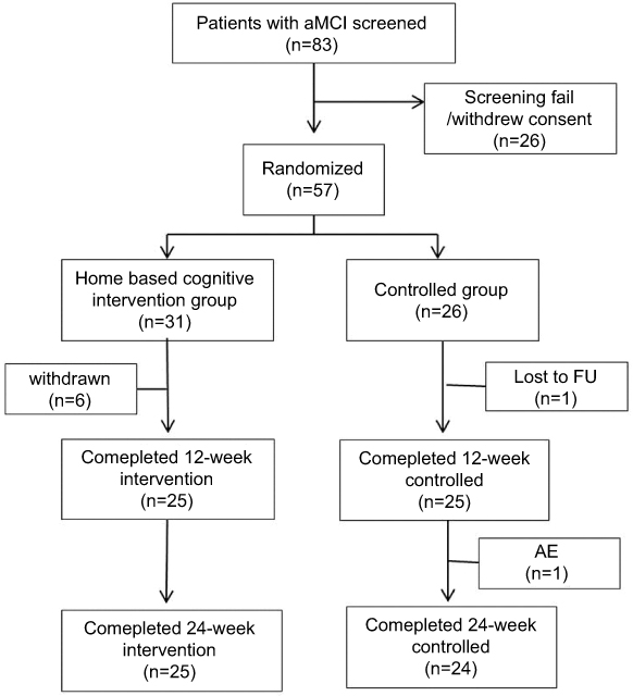 |
Figure 1 The flow diagram of enrollment.Abbreviations: FU, follow-up; AE, adverse event. |
Baseline characteristics, including age and sex, cognitive function, comorbidities and medication history, were not significantly different between groups (Table 1). In both groups, there were no significant differences in MMSE, CDR-SB, Modified ADAS-Cog score, or GDS-15 (SGDS-K).
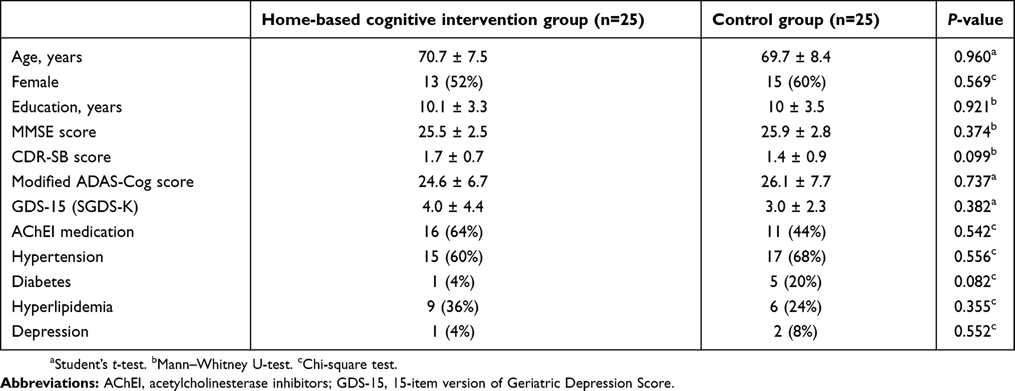 |
Table 1 Baseline characteristics and demographics of subjects |
Outcome of clinical symptoms
Changes in ADAS-Cog score for efficacy of cognitive training at 12 and 24 weeks are shown in Table 2. There was no significant difference versus baseline in the change in modified ADAS-Cog at 12 and 24 weeks between the control and training groups. However, ADAS-Cog score showed increased tendency at 24 weeks PI in the training group compared to control group. The trend of the change in ADAS-Cog score is depicted in Figure 2.
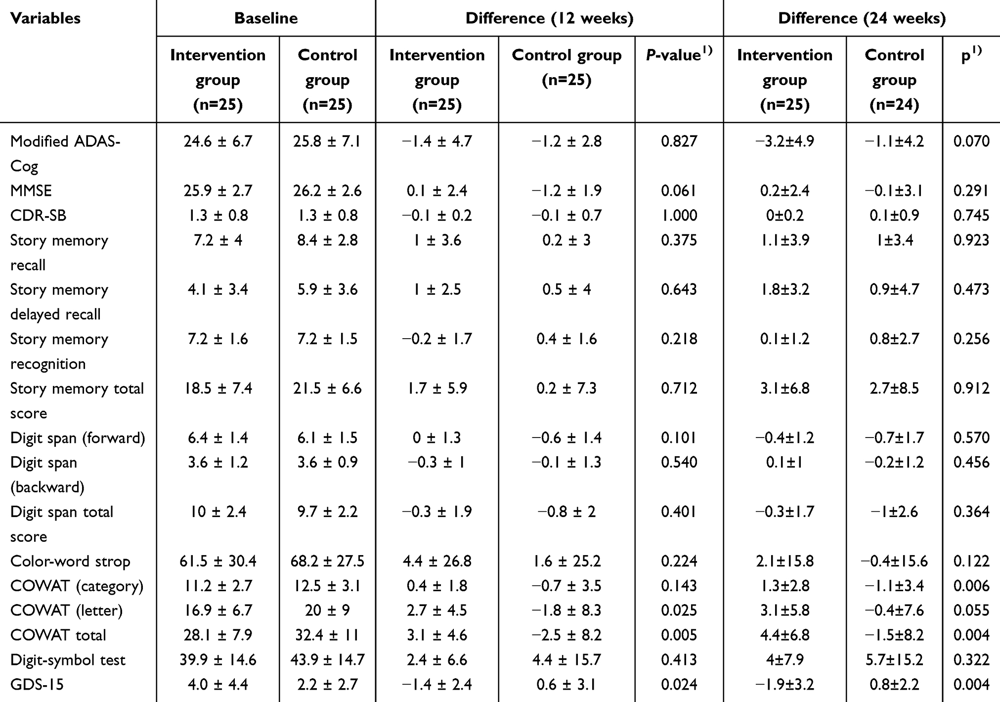 |
Table 2 Changes in efficacy outcomes at 12 and 24 weeks |
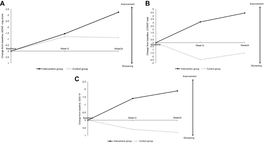 |
Figure 2 Changes from baseline at 12 weeks and 24 weeks on efficacy outcomes. Cognitive training significantly improved ADAS-Cog score at 24 weeks (A), COWAT total score at 12 and 24 weeks (B) and GDS-15 at 12 and 24 weeks (C). (A) Changes from baseline at 12 weeks and 24 weeks on the Modified ADAS-Cog. (B) COWAT (total) score change from baseline on Intervention group vs Control (C) GDS-15 (SGDS-K) score change from baseline on Intervention group vs Control.Abbreviation: COWAT, Controlled oral word association test. |
Controlled Oral Word Association Test (COWAT) total score, COWAT letter score, and GDS-15 scale were significantly improved in the training group compared with the control group at 12 weeks PI. At 24 weeks PI, COWAT total score and GDS-15 continued to improve in the training group versus control group. There was no significant difference in the change in other clinical scales including CDR-SB, story memory (recall, delayed recall, recognition, total score), digital span (forward, backward, total), color word strop and digital-symbol test at 12 and 24 weeks PI. MMSE score showed a tendency to improve in the training group even without statistical significance at 12 weeks.
Outcome of brain metabolism
The demographics, baseline cognitive function and comorbidity of participants who underwent PET/CT were not different among all participants (Table 3). There was a >20% difference in brain metabolism in one control-group patient, versus 10–20% in two patients in the training group. Other participants showed no significant changes (Figure 3).
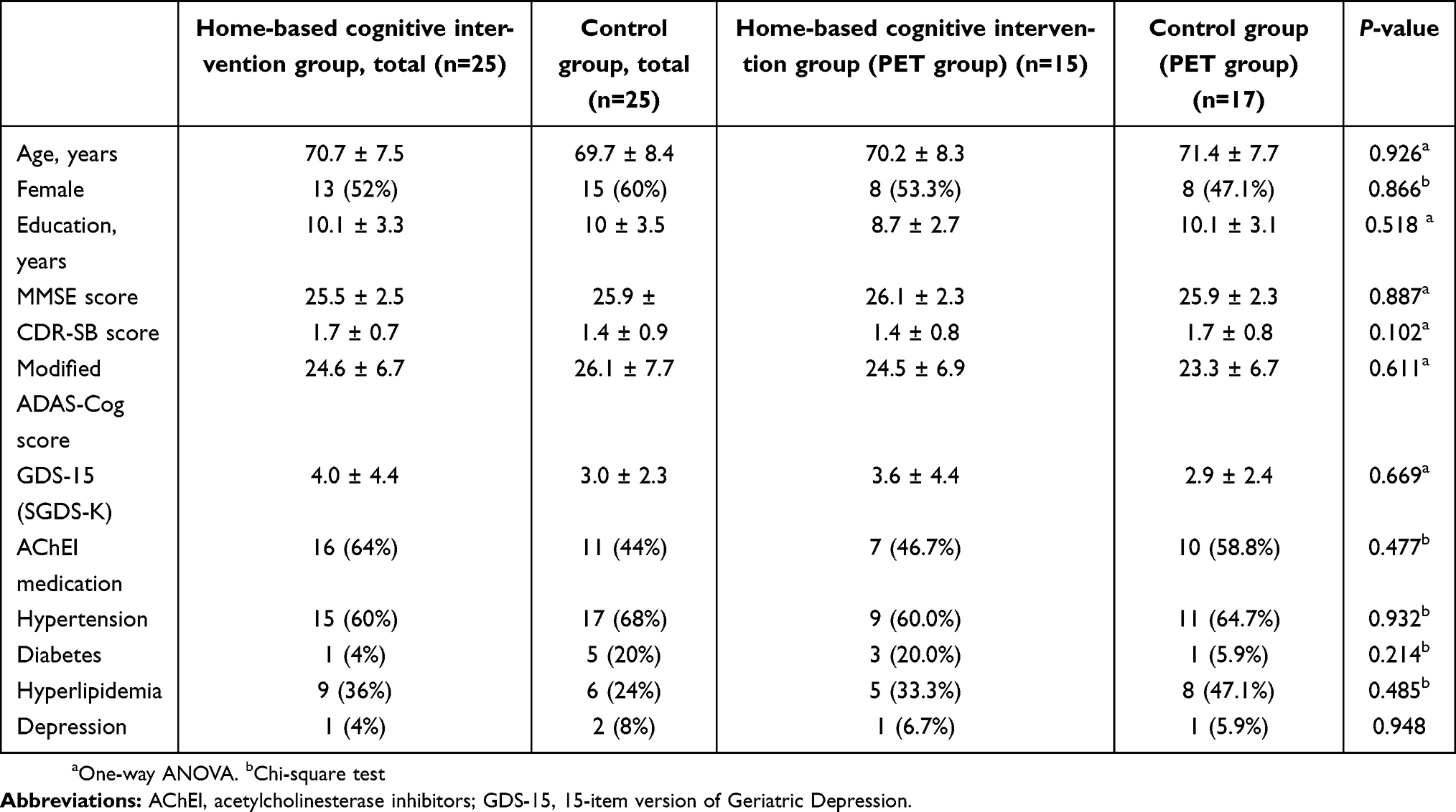 |
Table 3 The basic demographic features of participants who performed PET CT comparing all participants |
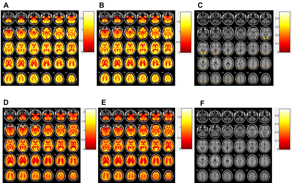 |
Figure 3 Distribution of SUVr before treatment (A), after treatment (B), and more than 20% increased region after treatment (C) in participant with change (Fig 3-1). Distribution of SUVr before treatment (D), after treatment (E), and no increased region after treatment (F) in participant without change (Fig 3-2). |
There was no significant difference in brain metabolism between the training and control groups. However, we found that the training group showed a tendency toward increased brain metabolism.
Discussion
This study evaluated the effect of HCI on cognition in MCI and brain metabolism. We found that HCI had a tendency to improve ADAS-Cog at 24 weeks from the beginning of cognitive training. Many studies have shown that cognitive training reduced cognitive decline in patients with MCI. In a meta-analysis, cognitive training significantly improved memory and subjective cognitive function compared with controls.18 Jeong et al reported that group- and home-based cognitive training, as used in this study, improved cognition in patients with amnestic MCI at 12 weeks.12 However, there was no significant improvement at the end of 12 weeks of cognitive training in our results. This disparity with the previous report may be due to small sample size and the limited local area. Our results suggest the possibility of a delayed effect of HCI on cognition. This finding is in accordance with the previous reports that the effect of cognitive training lasts up to 3 months.19
Our results showed that COWAT total score, used as a measurement of language and executive function, was improved at both 12 and 24 weeks. Our cognitive training program was intended to enhance many cognitive domains; language, executive function and depression were changed most prominently. There has been much evidence to support that each cognitive domain can be improved by specific cognitive therapy.20 However, inconsistent results have been reported regarding which cognitive domains were improved by cognitive training.21 This may be due to a lack of program standardization and bias of long-term training and follow-up. One meta-analysis, like our study, found that cognitive training improved memory and executive function. 18 Our results indicate that cognitive training is especially effective in executive function at 12 and 24 weeks.
We also found that HCI improved depressive mood in MCI at 12 and 24 weeks. Cognitive training for depression, which is usually called cognitive behavioral therapy, has also been widely published and clinically used.22,23
There were no significant differences in brain metabolism between the training and control groups. Some reports have investigated the functional changes in the brain after cognitive training; resting functional MRI showed that cognitive training increased functional connectivity in the hippocampus.24 FDG PET is known to monitor disease progression and previous reports of FDG PET imaging revealed that cognitive training attenuated the metabolic decline in cortical regions.14 We found a tendency toward increased glucose metabolism in the training group (Figure 3). We postulate several reasons for statistical insignificance: small sample size and heterogenic character of the MCI group; too short a time period to enable discrimination of changes in brain metabolism; and perhaps the inadequacy of FDG PET/CT as a tool for detecting minimal changes.25 New technology such as resting functional MRI and diffusion tensor imaging could be more accurate for our studies.
Although many studies on the effect of cognitive training in MCI patients have been published and the effect of our cognitive program has been proven in previous reports, our study has several unique aspects: first, our program was home-based, which can increase adherence.26 In Korea, most patients with dementia cannot receive non-pharmacological intervention because of the high cost and lack of infrastructure. HCI is self-directed learning, which can be easily assessed and enables long-term training.27 Furthermore, our results suggest that cognitive training has a delayed effect on cognition that lasts for 3 months after finishing the program. Second, we found that cognitive training improved scores for executive function and depression, which differs from previous studies.28 Finally, our study is a prospective trial investigating changes in brain metabolism after cognitive training in MCI patients. Although we failed to prove statistical significance, our results showed a high tendency toward increased brain metabolism. MCI is a neurodegenerative disease that is slowly progressive; therefore, 12 weeks for cognitive training may not be enough time to change brain metabolism. Previous reports for brain metabolism after cognitive training found that changes in FDG uptake persisted for 6 months.14 The small sample size imited statistical analysis; a postulate longer period or repetitive training with a larger sample size is required to determine the significance of changes observed.
Our study has several limitations. 1) We did not enroll MCI patients proven by amyloid PET imaging. This limitation might affect the result of glucose metabolism in FDG PET. 2) Our training program may not be well controlled compared with other studies because participants performed self-learning at home. Despite regular checking, there is individual variance in program performance. 3) Not all participants underwent FDG PET/CT.
Conclusion
We proved that HCI has an effect on frontal lobe function and depressive mood in patients with MCI. Cognitive training has a tendency to increase brain metabolism despite statistical significance. To better evaluate cognitive training and target cognitive domain, a well-designed, large prospective study with PET imaging is necessary.
Acknowledgments
This study was supported by a grant from the Original Technology Research Program for Brain Science through the National Research Foundation of Korea (NRF) funded by the Korean government (MSIP) [2014M3C7A1064752] and Eisai Korea Inc. The abstract of this paper was presented at the Alzheimer’s Association International Conference (AAIC) in Chicago (July 22–26, 2018) as a poster presentation with interim findings. The poster’s abstract was published in “Poster Abstracts” in Alzheimer’s & Dementia.
Disclosure
The authors report no conflicts of interest in this work.
References
1. Mufson EJ, Binder L, Counts SE, et al. Mild cognitive impairment: pathology and mechanisms. Acta Neuropathol. 2011;123(1):13–30. doi:10.1007/s00401-011-0884-1
2. Bahar-Fuchs A, Clare L, Woods B. Cognitive training and cognitive rehabilitation for mild to moderate Alzheimer’s disease and vascular dementia. Cochrane Database Syst Rev. 2013. doi:10.1002/14651858.CD003260.pub2
3. Massoud F, Gauthier S. Update on the pharmacological treatment of Alzheimer’s disease. Curr Neuropharmacol. 2010;8(1):69–80. doi:10.2174/157015910790909520
4. Karakaya T, Fußer F, Schröder J, Pantel J. Pharmacological treatment of mild cognitive impairment as a prodromal syndrome of Alzheimer´s disease. Curr Neuropharmacol. 2013;11(1):102–108.
5. Rakesh G, Szabo ST, Alexopoulos GS, Zannas AS. Strategies for dementia prevention: latest evidence and implications. Ther Adv Chronic Dis. 2017;8(8–9):121–136. doi:10.1177/2040622317712442
6. Woods B, Aguirre E, Spector AE, Orrell M. Cognitive stimulation to improve cognitive functioning in people with dementia. Cochrane Database Syst Rev. 2012;2:CD005562.
7. Keshavan MS, Vinogradov S, Rumsey J, Sherrill J, Wagner A. Cognitive training in mental disorders: update and future directions. Am J Psychiatry. 2014;171(5):510–522. doi:10.1176/appi.ajp.2013.13081075
8. Butler M, McCreedy E, Nelson VA, et al. Does cognitive training prevent cognitive decline? Ann Intern Med. 2017;168(1):63. doi:10.7326/M17-1531
9. Kallio EL, Ohman H, Kautiainen H, Hietanen M, Pitkala K. Cognitive training interventions for patients with Alzheimer’s disease: a systematic review. J Alzheimers Dis. 2017;56(4):1349–1372. doi:10.3233/JAD-160810
10. Jungwirth S, Zehetmayer S, Hinterberger M, Tragl KH, Fischer P. The validity of amnestic MCI and non-amnestic MCI at age 75 in the prediction of Alzheimer’s dementia and vascular dementia. Int Psychogeriatr. 2012;24(6):959–966. doi:10.1017/S1041610211002870
11. Buschert VC, Friese U, Teipel SJ, et al. Effects of a newly developed cognitive intervention in amnestic mild cognitive impairment and mild Alzheimer’s disease: a pilot study. J Alzheimers Dis. 2011;25(4):679–694. doi:10.3233/JAD-2011-100999
12. Jeong JH, Na HR, Choi SH, et al. Group- and home-based cognitive intervention for patients with mild cognitive impairment: a randomized controlled trial. Psychother Psychosom. 2016;85(4):198–207. doi:10.1159/000442261
13. Habeck C, Risacher S, Lee GJ, et al. Relationship between baseline brain metabolism measured using [18F]FDG PET and memory and executive function in prodromal and early Alzheimer’s disease. Brain Imaging Behav. 2012;6(4):568–583. doi:10.1007/s11682-012-9208-x
14. Forster S, Buschert VC, Buchholz HG, et al. Effects of a 6-month cognitive intervention program on brain metabolism in amnestic mild cognitive impairment and mild Alzheimer’s disease. J Alzheimers Dis. 2011;25(4):695–706. doi:10.3233/JAD-2011-100996
15. Andrea C, Maria Chiara G, Francesco B, Massimo Del S. FDG-PET in the evaluation of brain metabolic changes induced by cognitive stimulation in aMCI subjects. Curr Radiopharm. 2015;8(1):69–75.
16. Petersen RC, Aisen P, Boeve BF, et al. Mild cognitive impairment due to Alzheimer disease in the community. Ann Neurol. 2013;74(2):199–208. doi:10.1002/ana.23931
17. Schrag A, Schott JM. Alzheimer’s disease neuroimaging I. What is the clinically relevant change on the ADAS-Cog? J Neurol Neurosurg Psychiatry. 2012;83(2):171–173. doi:10.1136/jnnp-2011-300881
18. Kelly ME, Loughrey D, Lawlor BA, Robertson IH, Walsh C, Brennan S. The impact of cognitive training and mental stimulation on cognitive and everyday functioning of healthy older adults: A systematic review and meta-analysis. Ageing Res Rev. 2014;15:28–43. doi:10.1016/j.arr.2014.02.004
19. Jaeggi SM, Buschkuehl M, Jonides J, Shah P. Short- and long-term benefits of cognitive training. Proc National Acad Sci. 2011;108(25):10081–10086. doi:10.1073/pnas.1103228108
20. Nozawa T, Taki Y, Kanno A, et al. Effects of different types of cognitive training on cognitive function, brain structure, and driving safety in senior daily drivers: a pilot study. Behav Neurol. 2015;2015:525901. doi:10.1155/2015/534681
21. Edwards JD, Fausto BA, Tetlow AM, Corona RT, Valdés EG. Systematic review and meta-analyses of useful field of view cognitive training. Neurosci Biobehav Rev. 2018;84:72–91. doi:10.1016/j.neubiorev.2017.11.004
22. Johnsen TJ, Friborg O. The effects of cognitive behavioral therapy as an anti-depressive treatment is falling: A meta-analysis. Psychol Bull. 2015;141(4):747–768. doi:10.1037/bul0000015
23. Motter JN, Pimontel MA, Rindskopf D, Devanand DP, Doraiswamy PM, Sneed JR. Computerized cognitive training and functional recovery in major depressive disorder: a meta-analysis. J Affect Disord. 2016;189:184–191. doi:10.1016/j.jad.2015.09.022
24. Lampit A, Hallock H, Suo C, Naismith SL, Valenzuela M. Cognitive training-induced short-term functional and long-term structural plastic change is related to gains in global cognition in healthy older adults: a pilot study. Front Aging Neurosci. 2015;7:14. doi:10.3389/fnagi.2015.00014
25. Willis SL, Tennstedt SL, Marsiske M, et al. Long-term effects of cognitive training on everyday functional outcomes in older adults. Jama. 2006;296(23):2805. doi:10.1001/jama.296.23.2805
26. Shatil E, Metzer A, Horvitz O, Miller A. Home-based personalized cognitive training in MS patients: a study of adherence and cognitive performance. NeuroRehabilitation. 2010;26(2):143–153. doi:10.3233/NRE-2010-0546
27. Voerman JS, Klerk C, Merelle SY, et al. Long-term follow-up of home-based behavioral management training provided by migraine patients. Cephalalgia. 2014;34(5):357–364. doi:10.1177/0333102413515337
28. Smith GE, Housen P, Yaffe K, et al. A cognitive training program based on principles of brain plasticity: results from the Improvement in Memory with Plasticity-based Adaptive Cognitive Training (IMPACT) study. J Am Geriatr Soc. 2009;57(4):594–603. doi:10.1111/j.1532-5415.2008.02167.x
 © 2019 The Author(s). This work is published and licensed by Dove Medical Press Limited. The full terms of this license are available at https://www.dovepress.com/terms.php and incorporate the Creative Commons Attribution - Non Commercial (unported, v3.0) License.
By accessing the work you hereby accept the Terms. Non-commercial uses of the work are permitted without any further permission from Dove Medical Press Limited, provided the work is properly attributed. For permission for commercial use of this work, please see paragraphs 4.2 and 5 of our Terms.
© 2019 The Author(s). This work is published and licensed by Dove Medical Press Limited. The full terms of this license are available at https://www.dovepress.com/terms.php and incorporate the Creative Commons Attribution - Non Commercial (unported, v3.0) License.
By accessing the work you hereby accept the Terms. Non-commercial uses of the work are permitted without any further permission from Dove Medical Press Limited, provided the work is properly attributed. For permission for commercial use of this work, please see paragraphs 4.2 and 5 of our Terms.