")
Back to Journals » Drug Design, Development and Therapy » Volume 16
Efficacy of Nimodipine Combined with Latanoprost in Treating Open-Angle Glaucoma and Its Influence on Ocular Hemodynamics and Visual Field Defects
Received 7 December 2021
Accepted for publication 27 February 2022
Published 21 March 2022 Volume 2022:16 Pages 749—757
DOI https://doi.org/10.2147/DDDT.S352876
Checked for plagiarism Yes
Review by Single anonymous peer review
Peer reviewer comments 2
Editor who approved publication: Prof. Dr. Tin Wui Wong
Hui-Ping Duan,1 Rong Liu2
1Department of Ophthalmology, Shanxi Province Fenyang Hospital, Fenyang, 032200, Shanxi Province, People’s Republic of China; 2Department of Ophthalmology, Shanxi Datong University, Datong, 037000, Shanxi Province, People’s Republic of China
Correspondence: Hui-Ping Duan, Department of Ophthalmology, Shanxi Province Fenyang Hospital, Fenyang, 032200, Shanxi Province, People’s Republic of China, Tel +86-13037099450, Email [email protected]
Background: Open-angle glaucoma is a common ophthalmic disease, which has a great impact on the vision of middle-aged and elderly people. Medication plays an important role in the treatment of glaucoma, so finding effective drug treatment is of great significance to improve the quality of life of glaucoma patients.
Objective: To explore the curative effect of nimodipine combined with latanoprost in the treatment of open-angle glaucoma and its effect on ocular hemodynamics and visual field defects.
Methods: This study retrospectively analyzed the clinical data of 87 patients with open-angle glaucoma who came to the Shanxi Province Fenyang Hospital and The First Affiliated Hospital of Shanxi Datong University for treatment from January 2019 to January 2021. According to different treatment plans, the patients were divided into two groups: an observation group (n = 46) treated with nimodipine combined with latanoprost, and a control group (n = 41) treated by latanoprost monotherapy. Treatment efficacy, hemodynamics, visual field defects, 24-hour peak intraocular pressure, binocular optic disc parameters, adverse reactions and quality of life were recorded and compared between two groups of patients.
Results: The overall therapeutic effect of the observation group was significantly better than that in the control group. After treatment, ocular hemodynamics, visual field defects, 24-hour peak intraocular pressure, binocular optic disc parameters and life quality of both groups were obviously improved compared to those before treatment, with more significant improvements in the observation group. In addition, there was no significant difference in the incidence of adverse reactions between the two groups.
Conclusion: Nimodipine combined with latanoprost eye drops is effective in the treatment of primary open-angle glaucoma, which could effectively improve the ocular hemodynamics and visual field defects of patients with fewer adverse reactions and higher safety. Therefore, it can be further promoted and used in clinical practice.
Keywords: nimodipine, latanoprost, open-angle glaucoma, ocular hemodynamics, degree of visual field defects
Introduction
Open-angle glaucoma is a common ophthalmological disease with a predilection for middle-aged and elderly people. Due to the insidious condition and slow progression of the disease, patients often miss the optimal treatment timing without knowing it, resulting in optic neuropathy and even blindness; Therefore, it is particularly important to choose an appropriate treatment in clinical practice.1,2 The onset of open-angle glaucoma is closely associated with the metabolic imbalance of endogenous regulatory factors, increased resistance of aqueous humor outflow pathways, and abnormal metabolism of trabecular meshwork cells.3
The treatment of glaucoma is mainly to control intraocular pressure via multiple methods such as drug therapy, laser treatment and surgery.4 In recent years, many glaucoma patients choose surgery or laser treatment for the advancement of laser and surgical technology. However, as a common initial treatment and supplementary treatment after surgery or laser, drug therapy still plays an important role in the treatment of glaucoma.5,6 The drug treatment of glaucoma is mainly based on local eye drops. Latanoprost eye drops are a new type of phenyl-substituted propyl ester prostaglandin F2a, which is a selective F2a receptor agonist. Different from other anti-glaucoma drugs, it does not change the flow coefficient of aqueous humor, aqueous humor production and episcleral venous pressure, but significantly relieves the high intraocular pressure in patients with open-angle glaucoma and ocular hypertension by improving trabecular and uveoscleral drainage, thus achieving therapeutic effects. However, there are also some patients who failed to benefit from it.7 Nimodipine has the effect of relaxing vascular smooth muscle and reducing intracellular calcium ions, which is commonly used for the treatment of diseases such as hypertension and arrhythmia in clinical practice. In recent years, some scholars have found that nimodipine also has a certain protective effect on retinal ganglion cells.8,9 However, there are relatively few reports on the treatment of open-angle glaucoma with nimodipine combined with latanoprost.
Therefore, this study explores the efficacy and safety of nimodipine combined with latanoprost in the treatment of open-angle glaucoma, aiming to provide more clinical evidence for the treatment of open-angle glaucoma.
Materials and Methods
Clinical Information
A retrospective analysis was made on 87 patients with open-angle glaucoma who visited Shanxi Province Fenyang Hospital and The First Affiliated Hospital of Shanxi Datong University from January 2019 to January 2021. According to different treatment plans and options, 46 patients treated with nimodipine combined with latanoprost were included in the observation group and 41 patients treated with latanoprost monotherapy were included in the control group. Inclusion criteria: (1) Patients who meet the diagnostic criteria for primary open-angle glaucoma with intraocular pressure> 21mmHg, and diagnosed with glaucoma led by insufficient blood supply to the optic nerve head, or optic nerve fiber layer defect or visual field defects, with open chamber angle; (2) Patients with normal cornea as indicated by slit-lamp examination; (3) Patients with corrected vision> 0.3; (4) No use of carbonic anhydrase inhibitors, cholinergic inhibitors, or β-receptor blockers in the past two weeks before treatment; (5) Patients aged 40–55 years. Exclusion criteria: (1) Patients with a history of eye surgery; (2) Patients with pathological changes caused by improper medication; (3) Patients with angle closure or history of acute angle-closure glaucoma; (4) Patients complicated with other acute and chronic ocular diseases such as conjunctivitis, keratitis and uveitis; (5) Patients during pregnancy or lactation; (6) Patients who had a history of allergies to research drugs; (7) Patients with severe systemic diseases; (8) Patients with poor fixation and false negatives or false positives in visual field defect detection; (9) Patients with malignant tumors. All patients gave consent to the study with a written informed consent form signed. This study has been approved by the Ethics Committee of the Shanxi Province Fenyang Hospital and The First Affiliated Hospital of Shanxi Datong University and was conducted in accordance with the Declaration of Helsinki.
Treatment
Patients in the control group were treated with latanoprost eye drops (Lunanbeite Pharmaceutical Co., Ltd., SFDA Approval No. H20044234), 1 drop/time, once daily. On the basis of the control group, the observation group was given nimodipine tablets (Bayer Healthcare Co., Ltd., SFDA Approval No. H20003010) orally, 30 mg/time, once a day. The treatment lasted for 6 months in both groups.
Outcome Measures
- Therapeutic effects of patients in the two groups were evaluated and compared according to the following criteria: markedly effective: clinical symptoms basically disappeared, retinal circulation status was basically normal, visual acuity improved by ≥2 lines, and the visual field of 5 adjacent punctuation points expanded by ≥5°; effective: clinical symptoms improved, retinal circulation status improved, vision improved by 1 line, and visual field expansion below 5°; ineffective: clinical symptoms did not alleviate or aggravate, retinal circulation status did not improve, and vision was not improved or even worsened. Total effective rate = (number of markedly effective cases + number of effective cases)/total number of cases × 100%. (2) The color Doppler ultrasound system (SIEMENS, model ACUSON Oxana 3) was used to detect the ocular blood flow indicators of two groups of patients before and after treatment, including the peak systolic velocity (PSV), end diastolic velocity (EDV) of arteriae retinae centralis as well as vascular resistance index (RI). The probe frequency of the ultrasonic instrument was 7.5 Hz, and the linear array scanning was performed for 3–5 consecutive cardiac cycles. After confirming the best frequency spectrum, the image was frozen to measure PSV, EDV and RI. (3) Visual field defects: The degree of visual field defects before and after treatment was recorded, including the upper, below, temporal and nasal visual fields. (4) Pulsair intellipuff tonometer (Keeler, UK) was to detect and record the 24-hour peak intraocular pressure of two groups of patients before and 4 weeks after treatment. Patients were asked to take a sitting position for 3 consecutive measurements, and the average data taken. (5) Detection of optic disc parameters: The optic disc scanning mode in the optical coherence tomography (OCT) was selected for scanning and analysis, and the detection data of patients with dilated pupils were obtained. The data program that comes with OCT is then used for data processing and analysis, and the binocular optic disc parameters such as the vertical cup-to-disk ratio (C/D), average C/D, and rim area (RA) were automatically generated by the data program. (6) Adverse reactions: During treatment, the occurrence of adverse reactions such as arrhythmia, rapid heart rate, shortness of breath, and conjunctival congestion were observed in both groups. (7) The Chinese version of the National Eye Institute Visual Function Questionnaire-25 (NEI VFQ-25)10 was used to assess visual-related quality of life of patients. This scale includes 12 dimensions (26 items), including overall vision, near/distance activities, color vision, peripheral vision, mental health, etc. Responses in each dimension were divided into 6 levels (A, B, C, D, E, F), with a NEI VFQ-25 score ranging from 0 to 100. The higher the score, the less the damage and the better the quality of life.
Statistical Methods
SPSS 18.0 statistical software (Beijing Wangshu Times Technology Co., Ltd.) was used for statistical analysis of the data, and GraphPad Prism 6 (GraphPad Software) was used for image rendering. Count data, expressed as number of cases and percentages (%), were compared using the χ2 test. For measurement data, paired t-test was used for intra-group comparisons before and after treatment, and independent t-test for inter-group comparisons. Differences with P<0.05 were considered statistically significant.
Results
Comparison of General Information
Two groups of patients were comparable because of negligible differences in gender, age, smoking history, etc. (P>0.05) (Table 1).
 |
Table 1 General Information of Patients [n (%)] |
Comparison of Therapeutic Effects
The number of patients in the observation group with markedly effective, effective and ineffective treatment was 30, 13 and 3, respectively. And those of the control group were 18, 14, and 9, respectively. The total effective rate of the observation group was 93.48%, which was significantly higher than that of 78.05% in the control group (P<0.05) (Table 2).
 |
Table 2 Comparison of Therapeutic Effects Between the Two Groups [n (%)] |
Comparison of Ocular Hemodynamic Indexes Between the Two Groups
Before treatment, there was no significant difference in ocular hemodynamic indexes between the two groups (P>0.05). After treatment, PSV and EDV levels increased while RI level decreased significantly in the observation group compared with those before treatment. And the changes of the above hemodynamic indexes in the observation group were more obvious compared with the control group (P<0.05) (Figure 1).
 |
Figure 1 Comparison of ocular hemodynamic indicators between the two groups; (A) comparison of PSV between the two groups before and after treatment; (B) comparison of EDV between the two groups before and after treatment; (C) comparison of RI between the two groups before and after treatment. *Indicates P<0.05. |
Comparison of Visual Field Defects Between the Two Groups
Before treatment, no significant difference was observed in the degree of visual field defects between the two groups in all directions (P>0.05). After treatment, the range of visual field defects in both groups was significantly reduced, and that of the observation group was significantly smaller compared with the control group (P<0.05) (Figure 2).
 |
Figure 2 Comparison of visual field defects between the two groups; (A) comparison of upper visual field between the two groups before and after treatment; (B) comparison of lower visual field between the two groups before and after treatment; (C) comparison of temporal visual field between the two groups before and after treatment; (D) comparison of nasal visual field between the two groups before and after treatment. *Indicates P<0.05. |
Comparison of 24-Hour Peak Intraocular Pressure Before and After Treatment Between the Two Groups
Compared with before treatment, the 24-hour peak intraocular pressure of both groups decreased significantly after treatment (P<0.05); and the 24-hour peak intraocular pressure of the observation group was significantly better than that of the control group (P<0.05) (Table 3).
 |
Table 3 Comparison of 24-Hour Peak Intraocular Pressure Before and After Treatment Between the Two Groups |
Comparison of Binocular Optic Disc Parameters Between the Two Groups
There was no significant difference in vertical C/D, average C/D, visual field MD, and RA between the two groups before treatment (P>0.05). After treatment, the vertical C/D, average C/D, and visual field MD values of both groups decreased, while the RA value increased, with statistical significance (P<0.05). Compared with the control group, the optic disc parameters and visual field MD of the observation group were significantly better after treatment (P<0.05) (Figure 3).
 |
Figure 3 Comparison of binocular optic disc parameters between the two groups; (A) comparison of vertical C/D between the two groups before and after treatment; (B) comparison of average C/D between the two groups before and after treatment; (C) comparison of MD between the two groups before and after treatment; (D) comparison of RA values between the two groups before and after treatment. *Indicates P<0.05. |
Comparison of the Incidence of Adverse Reactions Between the Two Groups
The number of patients in the observation group who had arrhythmia, rapid heart rate, shortness of breath, and conjunctival congestion were 2, 1, 1, and 2, respectively, with an adverse reaction rate of 13.04%. And the corresponding number of cases in the control group were 1, 1, 0, and 2, respectively, with an incidence of adverse reactions of 9.76%. The two groups showed no significant difference in the incidence of adverse reactions (P>0.05) (Table 4).
 |
Table 4 Comparison of the Incidence of Adverse Reactions Between the Two Groups [n (%)] |
Comparison of Vision-Related Quality of Life Before and After Treatment Between the Two Groups
Compared with before treatment, the total score of NEI VFQ-25 increased significantly in both groups after treatment (P<0.05) and was higher in the observation group compared with the control group (P<0.05) (Table 5).
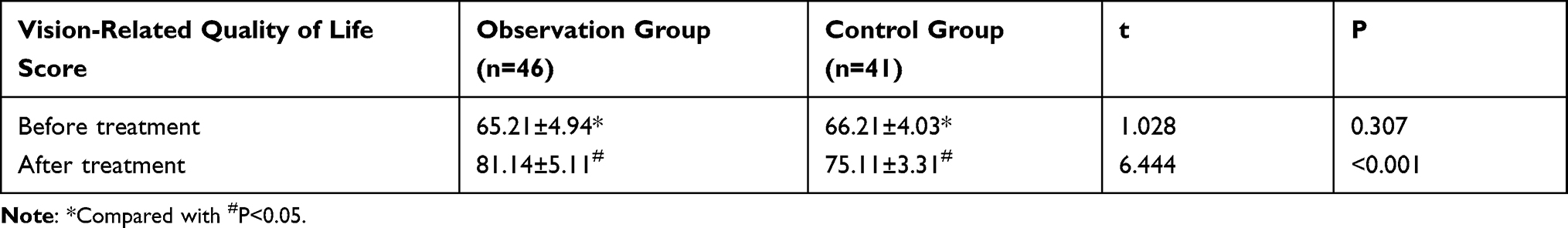 |
Table 5 Comparison of Vision-Related Quality of Life Scores Between the Two Groups Before and After Treatment |
Discussion
Open-angle glaucoma is characterized by continuous or intermittent increase in intraocular pressure, which compresses the optic papillary blood vessels and optic nerve fibers, resulting in insufficient blood supply to the optic nerve fiber bundles, impairment of visual function, visual field defects, and irreversible damage to the eye tissue, with a high blindness rate.11,12 At present, glaucoma is mainly treated with medication, surgery, laser. Although with certain therapeutic effects, these treatments are not conducive to the long-term prognosis of patients. Therefore, medication is becoming increasingly important. And how to effectively prevent and treat glaucoma has become the focus of social and clinical research and research.13
In this study, we first compared the efficacy of latanoprost monotherapy and latanoprost combined with nimodipine in the treatment of patients with open-angle glaucoma. It was found that the efficacy of latanoprost combined with nimodipine outweighted latanoprost monotherapy. Latanoprost is a drug commonly used for glaucoma and ocular hypertension in recent years. Its main active ingredient is prostaglandin analogues (which can be hydrolyzed into free acid after it penetrates into the anterior chamber. It acts on the ciliary muscle and the protease of the uveoscleral channel, which relaxes the ciliary muscle, widens the intermuscular space, and reduces the resistance of aqueous humor outflow to increase the aqueous humor through the uveoscleral outflow, thereby reducing intraocular pressure), which can help relieve clinical symptoms of patients; However, this treatment is ineffective in some patients.14,15 Nimodipine, a dihydropyridine calcium antagonist, is mainly used to improve blood circulation during the convalescent period of acute cerebrovascular disease and cerebral vasospasm caused by various reasons, with a protective effect on neurons of patients with vascular dementia. It can inhibit the influx of calcium ions and dilate blood vessels by acting on the vascular smooth muscle cell membrane, which can actively alleviate the ischemia and hypoxia ischemia and hypoxia.16 In recent years, nimodipine has been shown to be a promising topical drug for the treatment of glaucoma by analyzing its drug targets.17 However, there are still few reports on the application of nimodipine combined with latanoprost in the treatment of open-angle glaucoma.
The cause of glaucoma is closely related to the blood supply of the ocular arteries, often accompanied by decreased blood flow of optic nerve artery and increased ocular RI. Therefore, improving optic nerve blood supply is also of great significance in the clinical treatment of glaucoma.18 The results of this study showed that the EDV and PSV of the two groups were significantly increased after treatment, and the RI was significantly decreased, with more significant changes in the observation group, suggesting the treatment of open-angle glaucoma with latanoprost combined with nimodipine could more significantly improve the ocular hemodynamic indicators of patients than latanoprost monotherapy. In addition, the 24-hour peak intraocular pressure is an important basis for the diagnosis of glaucoma.19 NEI VFQ-25, with favorable validity and reliability, can comprehensively assess vision-related quality of life of patients with eye diseases, which is suitable for the diagnosis of common clinical eye diseases.20 Optic disc damage and visual field defects are the main pathological changes of open-angle glaucoma. Patients showed a significant increase in optic cup morphological parameters such as vertical C/D and average C/D, a significant decrease in RA, and glaucomatous damage in the visual field.21 In this study, the 24-hour peak intraocular pressure, vertical C/D, average C/D, and visual field MD values of patients in the observation group were significantly lower than those in the control group during the same period, and the total score of NEI VFQ-25 and RA values were significantly increased, indicating that the effect of nimodipine combined with latanoprost eye drops in the treatment of primary open-angle glaucoma is definite. This is also the first time that we have comprehensively analyzed the efficacy of nimodipine combined with latanoprost eye drops in the treatment of primary open-angle glaucoma, and confirmed that it could effectively improve the visual field defects in patients with the disease.
Previous studies have found that in the treatment of glaucoma, nimodipine can protect the visual field of patients. Since glaucoma patients are mostly caused by the dysfunction of optic nerve and blood vessel regulation, it is difficult for blood vessels to regulate the increased blood flow when the intraocular pressure rises. Meanwhile, patients with primary open-angle glaucoma often have abnormal hemorheology. Nimodipine can relax blood vessels by inhibiting calcium ions entering into vascular smooth muscle cells, thereby reducing arterial resistance, avoiding vasospasm, enhancing nerve blood supply, and eventually improving the visual field of patients.22,23 Moreover, nimodipine can correct retinal ganglion cells (RGCs). As the intraocular pressure increases, the load also increases in RGCs of glaucoma patients. Nimodipine can prevent calcium ions from flowing into cells, correcting “calcium overload” of cells, and stabilizing cell membranes, thereby protecting RGCs and preventing the progression of optic nerve defects,24 which explains our findings. Meanwhile, the drug side effects of the two groups of the present study were mainly mild ocular congestion, irregular heartbeat and other symptoms, without serious adverse events, indicating that the combination therapy had high safety.
In summary, nimodipine combined with latanoprost eye drops is effective in the treatment of primary open-angle glaucoma, as it can effectively improve the ocular hemodynamics and visual field defects in patients with fewer adverse reactions and higher safety, which is worthy of clinical promotion. However, this study also has certain shortcomings. First, due to the small sample size, the conclusions of this study need to be further verified. Second, the specific mechanism of nimodipine combined with latanoprost in open-angle glaucoma needs to be further analyzed. In the future, we will further carry out large-scale multi-center data studies and provide more data support for our conclusions in combination with relevant basic research.
Author Contributions
All authors contributed to data analysis, drafting or revising the article, have agreed on the journal to which the article will be submitted, gave final approval for the version to be published, and agree to be accountable for all aspects of the work.
Funding
This study did not receive any funding in any form.
Disclosure
The authors declare no competing interests in this work.
References
1. Bertaud S, Aragno V, Baudouin C, Labbe A. [Primary open-angle glaucoma]. Rev Med Interne. 2019;40(7):445–452. French. doi:10.1016/j.revmed.2018.12.001
2. Marshall LL, Hayslett RL, Stevens GA. Therapy for open-angle glaucoma. Consult Pharm. 2018;33(8):432–445. doi:10.4140/TCP.n.2018.432
3. Eliseeva NV, Churnosov MI. [Genome-wide studies of primary open-angle glaucoma]. Vestn Oftalmol. 2020;136(5):129–135. Russian. doi:10.17116/oftalma2020136051129
4. Khawaja AP, Viswanathan AC. Are we ready for genetic testing for primary open-angle glaucoma? Eye (Lond). 2018;32(5):877–883. doi:10.1038/s41433-017-0011-1
5. Yilmaz KC, Sur Gungor S, Ciftci O, Akman A, Muderrisoglu H. Relationship between primary open angle glaucoma and blood pressure. Acta Cardiol. 2020;75(1):54–58. doi:10.1080/00015385.2018.1549004
6. Schwab C, Paar M, Fengler VH, et al. Vitreous albumin redox state in open-angle glaucoma patients and controls: a pilot study. Int Ophthalmol. 2020;40(4):999–1006. doi:10.1007/s10792-019-01268-5
7. Garway-Heath DF, Crabb DP, Bunce C, et al. Latanoprost for open-angle glaucoma (UKGTS): a randomised, multicentre, placebo-controlled trial. Lancet. 2015;385(9975):1295–1304. doi:10.1016/S0140-6736(14)62111-5
8. Tong J, Li J, Zhang QS, et al. Delayed cognitive deficits can be alleviated by calcium antagonist nimodipine by downregulation of apoptosis following whole brain radiotherapy. Oncol Lett. 2018;16(2):2525–2532. doi:10.3892/ol.2018.8968
9. Sun J, Zheng J, Wang F, Zhang G, Wu J. Effect of hyperbaric oxygen combined with nimodipine on treatment of diffuse brain injury. Exp Ther Med. 2018;15(6):4651–4658. doi:10.3892/etm.2018.6045
10. Mangione CM, Lee PP, Gutierrez PR, et al. Development of the 25-item National Eye Institute Visual Function Questionnaire. Arch Ophthalmol. 2001;119(7):1050–1058. doi:10.1001/archopht.119.7.1050
11. Schuster AK, Wagner FM, Pfeiffer N, Hoffmann EM. Risk factors for open-angle glaucoma and recommendations for glaucoma screening. Ophthalmologe. 2021;118(Suppl 2):145–152. doi:10.1007/s00347-021-01378-5
12. Erb C, Predel HG. [Relevance of arterial hypertension in primary open-angle glaucoma]. Klin Monbl Augenheilkd. 2014;231(2):136–143. German. doi:10.1055/s-0033-1360331
13. Rivera JL, Bell NP, Feldman RM. Risk factors for primary open angle glaucoma progression: what we know and what we need to know. Curr Opin Ophthalmol. 2008;19(2):102–106. doi:10.1097/ICU.0b013e3282f493b3
14. Asrani S, Bacharach J, Holland E, et al. Fixed-dose combination of netarsudil and latanoprost in ocular hypertension and open-angle glaucoma: pooled efficacy/safety analysis of phase 3 MERCURY-1 and −2. Adv Ther. 2020;37(4):1620–1631. doi:10.1007/s12325-020-01277-2
15. Aref AA, Geyman LS, Zakieh AR, Alotaibi HM. Netarsudil and latanoprost ophthalmic solution for the reduction of intraocular pressure in open-angle glaucoma or ocular hypertension. Expert Rev Clin Pharmacol. 2019;12(12):1073–1079. doi:10.1080/17512433.2019.1701435
16. Wang W, Xu R, Yao M. [Ultraearly treatment on sudden deafness and the influence on superoxide dismutase and malonyldialdehyde of serum]. Zhonghua Er Bi Yan Hou Ke Za Zhi. 2002;37(4):274–276. Chinese.
17. Maria DN, Abd-Elgawad AH, Soliman OA, El-Dahan MS, Jablonski MM. Nimodipine ophthalmic formulations for management of glaucoma. Pharm Res. 2017;34(4):809–824. 10.1007/s11095-017-2110-x
18. Wang Y, Liao Y, Nie X. Comparative evaluation of Latanoprostene Bunod, Timolol Maleate, and latanoprost Ophthalmic Solutions to assess their safety and efficacy in lowering intraocular pressure for the management of Open-Angle Glaucoma. Clinics (Sao Paulo). 2020;75:e1874. doi:10.6061/clinics/2020/e1874
19. Lin Z, Huang S, Huang P, Li C, Chen Z, Zhong Y. Concordance of 24-h intraocular pressure curve in patients with untreated unilateral primary open-angle glaucoma. Exp Ther Med. 2018;16(2):1461–1469. doi:10.3892/etm.2018.6315
20. Karadeniz Ugurlu S, Kocakaya Altundal AE, Altin Ekin M. Comparison of vision-related quality of life in primary open-angle glaucoma and dry-type age-related macular degeneration. Eye (Lond). 2017;31(3):395–405. doi:10.1038/eye.2016.219
21. Siesky B, Wentz SM, Januleviciene I, et al. Baseline structural characteristics of the optic nerve head and retinal nerve fiber layer are associated with progressive visual field loss in patients with open-angle glaucoma. PLoS One. 2020;15(8):e0236819. doi:10.1371/journal.pone.0236819
22. Beidoe G, Mousa SA. Current primary open-angle glaucoma treatments and future directions. Clin Ophthalmol. 2012;6:1699–1707. doi:10.2147/OPTH.S32933
23. Araie M, Mayama C. Use of calcium channel blockers for glaucoma. Prog Retin Eye Res. 2011;30(1):54–71. doi:10.1016/j.preteyeres.2010.09.002
24. Luksch A, Rainer G, Koyuncu D, et al. Effect of nimodipine on ocular blood flow and colour contrast sensitivity in patients with normal tension glaucoma. Br J Ophthalmol. 2005;89(1):21–25. doi:10.1136/bjo.2003.037671
 © 2022 The Author(s). This work is published and licensed by Dove Medical Press Limited. The full terms of this license are available at https://www.dovepress.com/terms.php and incorporate the Creative Commons Attribution - Non Commercial (unported, v3.0) License.
By accessing the work you hereby accept the Terms. Non-commercial uses of the work are permitted without any further permission from Dove Medical Press Limited, provided the work is properly attributed. For permission for commercial use of this work, please see paragraphs 4.2 and 5 of our Terms.
© 2022 The Author(s). This work is published and licensed by Dove Medical Press Limited. The full terms of this license are available at https://www.dovepress.com/terms.php and incorporate the Creative Commons Attribution - Non Commercial (unported, v3.0) License.
By accessing the work you hereby accept the Terms. Non-commercial uses of the work are permitted without any further permission from Dove Medical Press Limited, provided the work is properly attributed. For permission for commercial use of this work, please see paragraphs 4.2 and 5 of our Terms.