")
Back to Journals » Research and Reports in Neonatology » Volume 14
Ethmocephaly, the Rarest Sub-Type of Holoprosencephaly: A Case Report
Authors Geta G , Mesfin T , Tsegaye M , Bobe T, Bikila B, Hailu FC, Dejene T
Received 25 October 2023
Accepted for publication 30 January 2024
Published 2 February 2024 Volume 2024:14 Pages 35—38
DOI https://doi.org/10.2147/RRN.S444777
Checked for plagiarism Yes
Review by Single anonymous peer review
Peer reviewer comments 3
Editor who approved publication: Dr Robert Schelonka
Girma Geta,1 Telila Mesfin,2 Mesfin Tsegaye,2 Tufa Bobe,3 Bonsa Bikila,1 Feleke Chefik Hailu,4 Tafese Dejene5
1Madda Walabu University, Goba General Hospital, Department of Midwifery, Goba, Oromia, Ethiopia; 2Madda Walabu University, Goba General Hospital, School of Medicine, Goba, Oromia, Ethiopia; 3Department of Obstetrics and Gynecology, Madda Walabu University, Goba Referral Hospital, Goba, Oromia, Ethiopia; 4Madda Walabu University, Goba General Hospital, Department of Nursing, Goba, Oromia, Ethiopia; 5Dire Dawa University, Department of Obstetrics and Gynecology, Dire Dawa, Ethiopia
Correspondence: Girma Geta, Tel +251920954580, Email [email protected]
Introduction: Holoprosencephaly results from incomplete cleavage of the forebrain during embryogenesis. Clinical phenotypes vary from mild to severe. We present a case of a liveborn infant with the most severe form of holoprosencephaly, ethmocephaly, which in most cases is incompatible with survival beyond the immediate newborn period.
Case: A 2000 gram, male infant was born to a 28-year-old gravida who was human immunodeficiency virus (HIV)-positive and receiving antiretroviral therapy. At birth the infant was lethargic and had poor respiratory effort with central and peripheral cyanosis. On physical examination, we found ocular hypotelorism, a 6 cm midline proboscis located above the eyes and absent nasal structures.
Conclusion: Early detection by the prenatal ultrasound examination is important pregnancy and birth planning and to anticipate expected outcomes. Further study is indicated to understand the contribution of HIV infection and its treatment to the development and severity of holoprosencephaly.
Keywords: ethmocephaly, holoprosencephaly, hypotelorism, proboscis
Introduction
Holoprosencephaly results from incomplete cleavage of the forebrain to form two distinct hemispheres during embryogenesis. It affects 1 in 10,000 live births, with females being affected more often than males.1
Ethmocephaly is the rarest sub-type of holoprosencephaly2,3 characterized by an undivided cerebrum and a single ventricle. Physical manifestations include two well-formed orbits with absent or hypoplastic globes, hypotelorism, low-set ears, and a mid-line facial abnormality with a central proboscis.3–6 Ethmocephaly occurs in 1 in 15,000 live births and in 1 of 250 abortuses; thus, most affected fetuses are abolished prenatally.3,7
First trimester ultrasonography can detect ethmocephaly. Hypotelorism and a single common cerebral ventricle are two ultrasonography indicators. Other investigations include chromosomal analysis by karyotyping, genetic mutation analysis using denaturing high-performance liquid chromatography, and multiplex polymer chain reaction.7,8
Recently, some cases of holoprosencephaly have also been reported among HIV-positive mothers with no clear link between maternal HIV status and holoprosencephaly.9 In addition to genetic problems, congenital infections, alcohol and tobacco intake during pregnancy, and diabetes mellitus have been reported to be holoprosencephaly risk factors in previous reports.7,10 We report a case of a newborn infant with ethmocephaly born to an HIV-positive gravida receiving anti-retroviral therapy in Southeast Ethiopia.
Case Presentation
We present a case of a 2000-gram male infant, born to a gravida III, Para II, 28-year-old HIV-positive gravida receiving highly active anti-retroviral therapy by spontaneous vaginal delivery. On presentation to Goba Referral Hospital, sonographic evaluation revealed a viable pregnancy at 34 weeks estimated gestation, polyhydramnios, a large right-side cystic intracranial mass, a single ventricle, and a bulb-shaped appearance of the thalami. At birth, the infant was lethargic, had central and peripheral cyanosis, and hypothermia with a measured temperature of 31.1°C. The respiratory rate was 72 breaths/min, heart rate of 155 beats/min, and oxygen saturation of 95% with an oropharyngeal airway and supplemental oxygen delivered via face mask. On clinical examination the sagittal, coronal, and lambdoid cranial sutures were easily separable. The head circumference was 34 cm and the anterior and posterior fontanels were 3 × 4 cm and 1.5 × 2 cm respectively. There was ocular hypotelorism (inner canthal distance was 2 cm) and both eyes were present with small, but reactive pupils. Both upper and lower eyelids appeared normal. We noted a 6 cm midline proboscis located above the globes of the eyes and absent nasal structures. Otherwise, both ears appeared normally set with typical pinnae and open external auditory canals. The hard and soft palates were intact and without cleft. Both the mandible and maxilla were well formed. There were no supernumerary digits of the hands and feet, and they appeared normal in alignment and size. There was no palpable defect, hair tuft lipoma, or hemangioma over the entire spinal vertebrae column. The external genitalia appeared normal (Figure 1).
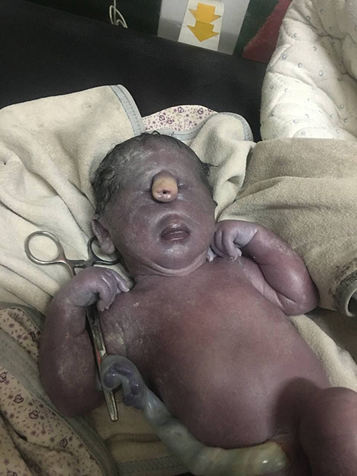 |
Figure 1 Clinical photograph showing ethmocephaly with ocular hypotelorism with a proboscis, and absent nasal structures. |
Initial stabilization of the child involved maintaining its temperature, airway, oxygenation, and providing dextrose-containing parenteral fluids. Despite providing all supportive treatments, the infant passed away at one hour of age following cardio-respiratory arrest. Genetic counseling was offered but required a trip abroad. The family could not afford the cost.
Discussion
Ethmocephaly is the rarest sub-type of holoprosencephaly with an undivided cerebrum, a single fused ventricle, a proboscis dividing the hypoteloric orbits, microphthalmos, low-set deformed ears, and absence of nasal bridge.3,6 Based on the phenotypic features, ethmocephaly can be distinguished from other holoprosencephaly conditions including cyclopia, in which the two eyes are fused in a single median orbit, and cebocephaly, which is characterized by a flattened snout with a single nostril below the level of the eye. The clinical findings of our case are similar to those reported by Das et al10 except for the finding of low set ears which we did not appreciate in our patient.
Most cases of ethmocephaly are believed to be sporadic without known causes. Environmental factors such as maternal alcoholism, prenatal exposure to teratogens, maternal insulin-dependent diabetes mellitus, and maternal infections may increase the risk for ethmocephaly.11 Ethmocephaly is associated with genetic factors, notably trisomy 13 or 18.12 In the case, we report HIV infection and the treatment may have contributed to the development of ethmocephaly. Sikakulya et al reported a case of a severe form of holoprosencephaly, cebocephaly, which has many overlapping clinical features with ethmocephaly, in a newborn born to an HIV-positive gravida in Eastern Uganda.9 Cebocephaly and ethmocephaly share a similar phenotype including dysplastic changes of the ethmoid bone and anterior portion of the sphenoid bone, with concomitant hypotelorism and defects of the medial orbital walls.6
Hypotelorism and a single common cerebral ventricle are two ultrasonography markers of ethmocephaly that can be detected in the first trimester.7,8 The birthing parent of the current case did have one antenatal visit at 12 weeks. We did not find records of any developmental abnormalities suspected during that visit. It is possible that during the only prenatal visit that even if an ultrasound was performed, it may not have been accurately interpreted.
Fetal assessment during antenatal visits is important for pregnancy and birth management decisions.5,9 In the present case, the diagnosis of holoprosencephaly was suspected just prior to birth. Termination of pregnancy was not an option, and there was little time for the family to prepare for the birth of a child with atypical brain development and little chance of surviving beyond the newborn period. Because of resource limitations, genetic studies and cranial imaging were not obtained after birth to identify risk factors other than the potential risk factor of in utero exposure to HIV and the medications to treat it.
Conclusion
Ethmocephaly carries a poor prognosis, and in most cases it is incompatible with life. Early detection by the prenatal ultrasound examination is important pregnancy and birth planning and to anticipate expected outcomes. Further study is indicated to understand the contribution of HIV infection and its treatment to the development and severity of holoprosencephaly.
Availability of Supporting Data
Data on the case clinical information informed consent forms and images are available for review from the corresponding author upon request.
Ethical Approval
No ethical approval is required for this case report.
Consent Form
Written informed consent was obtained from the parents of the case for publication of this case report and any accompanying images. A copy of the written consent is available for review by the Editor-in-Chief of this journal.
Disclosure
The authors declare that there are no conflicts of interest in this work.
References
1. Ariyo IJ, Mchaile DN, Magwizi M, et al. Alobar holoprosencephaly with cebocephaly in a neonate: a rare case report from Northern Tanzania. Internat J Surg Case Rep. 2022;93:106960. doi:10.1016/j.ijscr.2022.106960
2. Spirt BA, Oliphant M, Gordon LP. Fetal central nervous system abnormalities. Radiol Clin North Am. 1990;28(1):59–73. doi:10.1016/S0033-8389(22)01220-9
3. Dewan P, Rohatgi S, Roy S, et al. Ethmocephaly: a rare cephalic disorder. J Pediatr Neurosci. 2016;11(1):92–93. doi:10.4103/1817-1745.181262
4. Al-Shaqsi S, Al-Bulushi T, Al-Hinai Q. Proboscis lateralis: a case report of a rare giant craniofacial teratoma in an infant. Arch Plast Surg. 2018;45(6):578–582. doi:10.5999/aps.2017.01739
5. Winter TC, Kennedy AM, Woodward PJ. Holoprosencephaly: a survey of the entity, with embryology and fetal imaging. Radiographics. 2015;35(1):275–290. doi:10.1148/rg.351140040
6. Souza JP, Siebert JR, Beckwith JB. An anatomic comparison of cebocephaly and ethmocephaly. Teratology. 1990;42(4):347–357. doi:10.1002/tera.1420420404
7. Chatterjee R, Jain KC. Ethmocephaly. Indian Pediatr. 2001;38(10):1194.
8. Sivanathan J, Thilaganathan B. Book: genetics for obstetricians and gynaecologists: chapter: genetic markers on ultrasound scan. Best Pract Res Clin Obstet Gynaecol. 2017;42:64–85. doi:10.1016/j.bpobgyn.2017.03.005
9. Sikakulya FK, Kiyaka SM, Masereka R, et al. Alobar holoprosencephaly with cebocephaly in a neonate born to an HIV-positive mother in Eastern Uganda. Case Rep Otolaryngol. 2021;2021:1.
10. Das G, Das D, Das G, et al. Ethmocephaly with amniotic band syndrome. Middle East Afr J Ophthalmol. 2012;19(4):429–431. doi:10.4103/0974-9233.102769
11. Croen LA, Shaw GM, Lammer EJ. Risk factors for cytogenetically normal holoprosencephaly in California: a population-based case-control study. Am J Med Genet. 2000;90(4):320–325. doi:10.1002/(SICI)1096-8628(20000214)90:4<320::AID-AJMG11>3.0.CO;2-8
12. Roessler E, Muenke M. Holoprosencephaly: a paradigm for the complex genetics of brain development. J Inherit Metab Dis. 1998;21(5):481–497. doi:10.1023/A:1005406719292
 © 2024 The Author(s). This work is published and licensed by Dove Medical Press Limited. The full terms of this license are available at https://www.dovepress.com/terms.php and incorporate the Creative Commons Attribution - Non Commercial (unported, v3.0) License.
By accessing the work you hereby accept the Terms. Non-commercial uses of the work are permitted without any further permission from Dove Medical Press Limited, provided the work is properly attributed. For permission for commercial use of this work, please see paragraphs 4.2 and 5 of our Terms.
© 2024 The Author(s). This work is published and licensed by Dove Medical Press Limited. The full terms of this license are available at https://www.dovepress.com/terms.php and incorporate the Creative Commons Attribution - Non Commercial (unported, v3.0) License.
By accessing the work you hereby accept the Terms. Non-commercial uses of the work are permitted without any further permission from Dove Medical Press Limited, provided the work is properly attributed. For permission for commercial use of this work, please see paragraphs 4.2 and 5 of our Terms.