Back to Journals » Clinical Ophthalmology » Volume 3
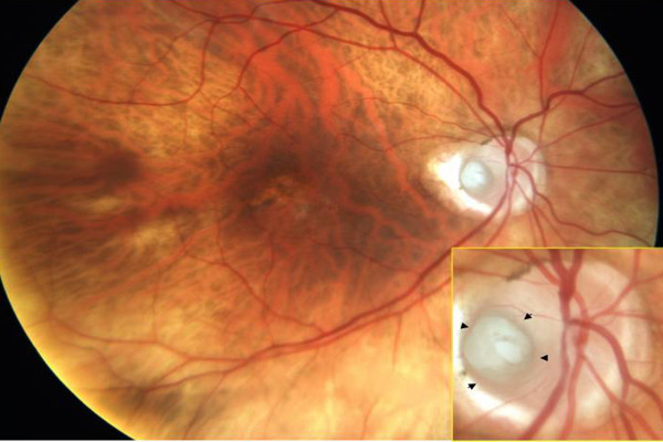
Functional microperimetry and SD-OCT confirm consecutive retinal atrophy from optic nerve pit
Authors Brar VS, Murthy RK, Chalam K
Published 11 November 2009 Volume 2009:3 Pages 625—628
DOI https://doi.org/10.2147/OPTH.S7908
Review by Single anonymous peer review
Peer reviewer comments 3

Vikram S Brar, Ravi K Murthy, K V Chalam
University of Florida College of Medicine, Department of Ophthalmology, Jacksonville, FL, USA
Abstract: A congenital anomaly, optic nerve pit is often associated with serous retinal detachment involving macula. Long standing serous detachment leads to outer retinal atrophy and decrease in visual sensitivity. Recently, spectral-domain optical coherence tomography (OCT) has been reported to demonstrate a communication between the optic nerve sheath and the subretinal space. Vitreous cavity is proposed as an alternate source of fluid for accumulation in the subretinal space. We imaged a patient with optic nerve pit with Spectralis OCT and report the findings seen including the presence of an area of peripapapillary retinal atrophy, due to the spontaneous resolution of associated long-standing retinal detachment.
Keywords: optic nerve pit, SD-OCT, autoflourescence, microperimetry
 © 2009 The Author(s). This work is published and licensed by Dove Medical Press Limited. The full terms of this license are available at https://www.dovepress.com/terms.php and incorporate the Creative Commons Attribution - Non Commercial (unported, v3.0) License.
By accessing the work you hereby accept the Terms. Non-commercial uses of the work are permitted without any further permission from Dove Medical Press Limited, provided the work is properly attributed. For permission for commercial use of this work, please see paragraphs 4.2 and 5 of our Terms.
