")
Back to Journals » Clinical, Cosmetic and Investigational Dermatology » Volume 15
The Application of Color Doppler Ultrasound in the Evaluation of the Efficacy of 595-nm Pulsed Dye Laser Combined with 755-nm Long-Pulse Alexandrite Laser in the Treatment of Hybrid Infantile Hemangiomas
Authors Lu Y , Zhou F, Gao Y, Jin W
Received 26 October 2022
Accepted for publication 14 December 2022
Published 22 December 2022 Volume 2022:15 Pages 2831—2839
DOI https://doi.org/10.2147/CCID.S393962
Checked for plagiarism Yes
Review by Single anonymous peer review
Peer reviewer comments 4
Editor who approved publication: Dr Jeffrey Weinberg
Yuwen Lu, Fangyan Zhou, Yu Gao, Wanwan Jin
Department of Dermatology, The Second Affiliated Hospital and Yuying Children’s Hospital of Wenzhou Medical University, Wenzhou, People’s Republic of China
Correspondence: Yu Gao; Wanwan Jin, Email [email protected]; [email protected]
Purpose: We used color Doppler ultrasound to conduct an objective evaluation of the 595-nm Pulsed Dye Laser (PDL) combined with 755-nm long-pulse alexandrite sequential laser treatment for hybrid IH.
Patients and Methods: A total of 116 cases of hybrid IH were selected for this study. The interval between laser treatments was around 4 weeks, and 6 laser treatments or complete removal of the tumor was the end point. All children underwent color Doppler ultrasonography at the 0th, 1st, 6th months of treatment. Children were grouped by gender, age (< 6 months, ≥ 6 months), thickness (< 8 mm, ≥ 8 mm), and location (face and neck, trunk, and extremities). Calculate the volume of IHs according to color Doppler ultrasound. The volume ratio before and after treatment was defined as the A-value. Treatment outcomes were defined as effective when the A-value < 75%.
Results: In total, 74 cases (63.79%) had effective outcomes. Overall, the samples showed a statistically significant difference in the reduction of IH volume after 6 months of laser treatment (P < 0.001). The treatment of < 6 months group had better efficacy than the ≥ 6 months group (P < 0.001), the treatment of thickness < 8 mm group had better efficacy than the thickness ≥ 8 mm group (P < 0.001) and there was no significant difference in efficacy between the three different location groups (P > 0.05). Greater reduction in blood flow in the group with the effective outcome than in the group with the ineffective outcome (P < 0.001).
Conclusion: Color Doppler ultrasound can be applied to the diagnosis of hybrid IH and to the evaluation of treatment time and outcomes, and it can help clinicians recognize hybrid IH with greater accuracy. The earlier intervention for hybrid IH we perform, the better outcomes will be.
Keywords: hybrid infantile hemangioma, color Doppler ultrasound, treatment, pulsed dye laser, alexandrite laser
Introduction
Infantile hemangioma (IH) is one of the most common benign vascular tumors in children. Statistically, the incidence of IH ranges from 2% to 10%.1 There has been a significant and steady increase in the incidence of IH over the past 30 years, attributing to the increasing incidence of prematurity and low birth weight.1,2 The growth of IH is not linear, most IH do not generally exist at birth, but appear within 1 month after birth and then proliferate. The first 6 months after birth, especially the first 3 months, is the early proliferative phase of IH, and the late proliferative phase is 6–9 months after birth, while a few IH may continue to proliferate until 2 years after birth.3 The regression phase is slower and may take 3–10 years.4 Current clinical treatments for IH include systemic pharmacotherapy, topical pharmacotherapy, laser therapy, surgical, and injection therapy.
In laser therapy, pulsed dye laser (PDL) is the most common device to treat IH. Because of its wavelength characteristics, PDL has high absorption coefficients for oxyhemoglobin (OHB) and deoxyhemoglobin (DHB). It has been extensively used to treat small IH lesions early, or to solve residual lesions during the regression phase. Due to the penetration depth of PDL, which is only 1–2 mm, it has limited effectiveness in treating thicker IH and may not be able to stop the proliferative growth of deeper IH.5 In numerous studies, the 755-nm long-pulse alexandrite laser, penetration depth 50–75% deeper than the PDL, has been proven safe and effective in the treatment of deeper IH.6,7
The clinical presentation of IH depends on its depth. The superficial IH is located in the superficial dermis and appears as a red lobulated plaque. The deep IH is located in the deep dermis and/or subcutaneous tissues and appears as a greenish-blue subcutaneous mass. The hybrid IH is located in both layers of the dermis and partially also includes subcutaneous tissues and exhibits clinical features that combine both of these categories.8
IH is typically diagnosed by the clinician’s subjective visual observation. Color Doppler ultrasound is a convenient, practical and non-invasive examination instrument that has become a routine diagnostic method for IH. It diagnoses various types of IH, measures the diameter, thickness and transverse diameter of the lesion, and shows internal echo and blood flow signal of the mass. It allows clinicians to choose the best treatment modality based on the depth and thickness of the IH and determine when to discontinue treatment.9,10
Therefore, in this study, we objectively evaluate the efficiency of 595-nm PDL combined with 755-nm long-pulse alexandrite sequential laser treatment for hybrid IH with the regular color Doppler ultrasound.
Materials and Methods
Study Design
This is a retrospective study in which children with hybrid infantile hemangioma who visited the dermatology department of the Second Affiliated Hospital and Yuying Children’s Hospital of Wenzhou Medical University for treatment from August 1, 2019 to August 1, 2022 were selected. This study complies with the Declaration of Helsinki and was approved by the ethics committee of the Second Affiliated Hospital and Yuying Children’s Hospital of Wenzhou Medical University (2022-K-126-01), and informed consent was obtained from the guardians of all recruited children.
Patients
The inclusion criteria for this study were as follows: 1. hybrid with low or medium risk infantile hemangiomas that meet the relevant diagnostic criteria of the Diagnostic and Treatment Guidelines of Hemangioma and Malformation of Vessels, 2019 edition;11 2. solitary infantile hemangiomas within 1 year of age; 3. first visit to our hospital, without any previous intervention; 4. children who underwent regular color Doppler ultrasound examinations during the treatment; 5. children who have been treated continuously for 6 months.
Exclusion criteria were as follows: 1. infantile hemangiomas that have been treated with topical or/and oral ß-adrenergic receptor inhibitors, lasers, injections and surgery in the past; 2. infantile hemangiomas with PHACE, LUMBAR, and other hemangioma and vascular malformation-related syndromes; 3. infantile hemangiomas with medium or high-risk features prone to ulceration, highly disfiguring damage, or functional impairment; 4. children with severe organic pathologies (eg, cardiopulmonary disorders); 5. children whose treatment regimen changed during the 6 months study period.
Treatment Options
Basic information about the children was gathered during the first visit, such as age, birth weight, gender and color Doppler ultrasound of IH. The interval between laser treatments was around 4 weeks, and 6 laser treatments or complete removal of the tumor was the end point. The Vbeam 595-nm pulsed dye laser (Candela Laser, Inc.) and 755-nm long-pulse alexandrite laser (Candela Laser, Inc.) were used to treat the patients sequentially based on their specific conditions. 595-nm PDL was applied with the following settings: wavelength 595 nm, energy density 6.0–7.0J/cm2, spot diameter 5–10mm, pulse width 0.45–20ms, dynamic cooling device (DCD) synchronous dynamic cooling system for 30–40ms jets and 20–30ms intervals. 755-nm long-pulse alexandrite laser was applied with the following settings: wavelength 755 nm, pulse width 3 ms, energy density 45–60 J/cm2, spot diameter 6–8 mm, DCD synchronous dynamic cooling system with 20 ms injection and 20 ms interval; cold compresses were applied for 15–30 minutes after laser treatment to reduce post-laser pain and eliminate swelling. Fusidic acid cream (Bright Future) was then applied topically for 7 days in order to prevent infection. Moreover, photographs of the lesions were taken with a camera (Canon PowerShot G11) before each treatment for efficiency observation.
Ultrasonography
The Yum MyLab Twice eHD color Doppler ultrasound diagnostic instrument with LA-523 line array probe at 4–13 MHz was used to evaluate children’s IH at the 0th (before laser treatment), 1st (after one laser treatment) and 6th months of the treatment. Color Doppler ultrasound was performed when the child was in a quiet state. If necessary, sedation (10% chloral hydrate enema, 0.5–1.0 mL/kg/dose) could be used for the child. The child received color Doppler ultrasound after falling asleep for 20 minutes. During ultrasonography, the type (superficial, deep or hybrid), size (diameter, thickness and transverse diameter), morphology, internal echogenicity, depth from the body surface and color Doppler flow imaging (CDFI) of the IH were determined.
Efficacy Evaluation
The estimated volume of the IH was diameter *thickness *transverse diameter * (π/6).12 The effect of the treatment was determined according to the size of the IH measured by color Doppler ultrasound. The volume ratio before and after treatment was defined as the A-value. Treatment results were categorized into Grade 1 (A-value ≥100%), Grade 2 (75% ≤ A-value < 100%) and Grade 3 (A-value <75%). As a criterion for clinical efficacy evaluation, treatment outcomes were defined as effective (Grade 3) and ineffective (Grades 1 and 2). CDFI’s outcomes were defined as reduced blood flow when the blood flow signal changed from hypervascularity to hypo vascularity or when the blood flow signal decreased by more than 50% compared with the first visit.
Side Effects
Parents were informed to observe and photograph the child’s IH changes regularly after the laser and were asked about the occurrence of local adverse events (eg, erythema, blister, scab, erosion, ulceration, scar, hyperpigmentation and hypopigmentation) and systemic adverse events (eg, asthma, bradycardia, hypotension, hypoglycemia, gastrointestinal disorders, sleep disorders and diarrhea) at each clinic visit. All adverse events were documented in detail.
Statistical Analysis
All statistical analyses were performed using the MacOS version of SPSS 26.0 (IBM Corp., Armonk, NY) software for data analysis. Normality testing was performed using the Shapiro–Wilk method. Samples that conformed to a normal distribution were analyzed using the independent samples t-test; samples that did not conform to a normal distribution were analyzed using the paired Wilcoxon signed-rank test or Kruskal–Wallis H-test; and the chi-square test or Fisher's exact test was performed for count variables. P < 0.05 was considered statistically significant.
Results
A total of 116 children with hybrid IH were included in this study (Table 1). There were 56 males (48.28%) and 60 females (51.72%), and the ratio of male to female was 1:1.07; 68 cases (58.62%) in the <6 months group and 48 cases (41.38%) in the ≥6 months group; a total of 46 cases (39.66%) in the face and neck region, 38 cases (32.76%) in the trunk region, and 32 cases (27.59%) in the extremity region. Meanwhile, the hybrid IH thickness measured by color Doppler ultrasound before treatment was divided into two groups, with thickness <8 mm defined as the shallower group and thickness ≥8 mm defined as the deeper group, with 72 cases (62.07%) in the shallower group and 44 cases (37.93%) in the deeper group.
 |
Table 1 Baseline Characteristics of the Children with Hybrid IH |
The A-value of the total sample after 1 time laser treatment was 95.94% ± 13.1% (x ± SD), and 30.17% of the children had an increase in volume compared to the pre-treatment period. The A-value of the overall sample after 6 months of sequential laser treatment was 64.68% ± 24.86%. In total, 74 children had Grade 3 (63.79%), 38 children had Grade 2 (32.76%), and 4 children had Grade 1 (3.45%). A total of 74 cases had an effective outcome (Figure 1), including 42 cases with color Doppler ultrasound suggestive of decreased blood flow and 32 cases with unchanged blood flow; 42 cases had an ineffective outcome, including 8 cases with decreased blood flow and 34 cases with unchanged blood flow. Among them, 4 children with increased volume were treated with oral propranolol or local sclerotherapy injection at the end of the study, and all achieved effective relief.
 |
Figure 1 Images of the lesions and color Doppler ultrasound images of a child with hybrid IH with a effective outcome at the 0th, 1st, 6th months. (A–C) At the 0th, 1st, 6th months, the images of the hybrid IHs. (D–F) At the 0th, 1st, 6th months, the diameters, depths and thicknesses in the ultrasound. (G–I) At the 0th, 1st, 6th months, the transverse diameters in the ultrasound. (J–L) At the 0th, 1st, 6th months, the CDFIs in the ultrasound. |
The difference in IH’s volumes before and after 1 time laser treatment in the overall sample was not statistically significant (P = 0.418>0.05). The overall sample showed a statistically significant difference in the reduction of IH’s volume after 6 months of laser treatment compared to pre-treatment (P < 0.001). The difference in efficacy between the different gender groups was not statistically significant (P = 0.468>0.05). The treatment was more effective in the <6 months group, with a statistically significant difference (χ2 = 22.837, P < 0.001) (Table 2). The efficacy of treatment in the shallower group was superior to that of the deeper group, with a statistically significant difference (χ2 = 16.260, P < 0.001) (Table 3). There was no statistically significant difference in efficacy among the three different location treatment groups (P = 0.400>0.05). More blood flow reduction is in the group with effective outcomes than in the group with ineffective outcomes, with a statistically significant difference (χ2 = 15.535, P < 0.001) (Table 4).
 |
Table 2 Efficacy After 6 Months of Laser Treatment of Age-Group |
 |
Table 3 Efficacy After 6 Months of Laser Treatment of Thickness-Group |
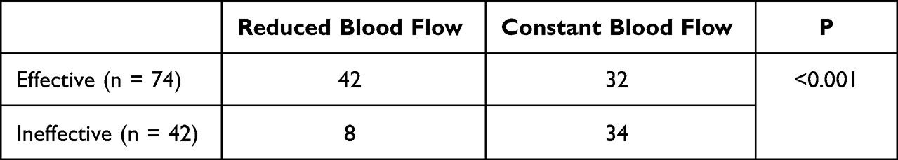 |
Table 4 The Relationship Between Outcomes and CDFI Changes |
None of the children in this study had systemic adverse events. In all, 28 children (24.14%) had adverse reactions, 9 had hyperpigmentation, 7 had hypopigmentation, 2 had scar, and 12 had blisters. One of them had blister combined with scar and one had hyperpigmentation combined with scar (Table 5). Children with hyperpigmentation and hypopigmentation showed the basic recoveries within 3 months of the follow-up after the end of the last treatment. 2 children with scars did not resolve significantly during the follow-up period after the end of treatment. Among the 12 children with blisters, excluding the 2 children with combined scarring or hyperpigmentation, the remaining 10 cases had only temporary skin lesions, and the blisters resolved after 1 week of topical fusidic acid cream.
 |
Table 5 Adverse Reactions During Treatment |
Discussion
This study retrospectively analyzed the efficacy of 595-nm PDL combined with 755-nm long-pulse alexandrite laser on 116 cases of hybrid IH after 6 months of sequential treatment in our dermatology department and was objectively reflected by color Doppler ultrasound. IH may occur everywhere on the body but primarily on the head, face and neck.13 There is a female predominance in the development of IH, and the male–female ratio is generally considered to be 1:2 to 1:5.14 In this study, the face and neck region predominated, consistent with the epidemiologically suggested results. The male–female ratio was nearly 1:1 in this study, which may be linked to the small sample size.
Oral propranolol is considered the current treatment of choice for IH, but it is not an ideal treatment for localized, solitary IH due to the side effects of propranolol, such as: hypoglycemia and cardiac burden.15 Topical ß-blockers, on the other hand, can only act on superficial IH due to the limitations of topical medications. In 1983, Anderson et al16 discovered that the theory of selective photothermolysis and laser therapy for the treatment of limited thicker IH had become a popular research direction over the past few years. Studies have shown that plasma concentrations of vascular endothelial growth factor (VEGF), a growth factor that can promote vascular proliferation, can be significantly reduced after laser treatment, with ultrasound suggesting reduced blood flow.17 However, many children with IH are not sensitive to laser treatment.18,19 In this study, four children increased in volume after laser treatment, and they were treated with oral propranolol or local sclerotherapy injections, which provided effective relief. Therefore, laser treatment can combine with other treatment modalities to obtain better results for IH. Asilian et al18 also demonstrated that timolol combined with 595-nm PDL had better efficacy and shorter treatment duration than 595-nm PDL alone.
The high absorption coefficient of OHB and DHB is one of the mechanisms of PDL in the treating IH, and PDL allows rapid resolution of residual lesions at the degenerative stage. Kessels et al20 randomized infants with IH into two groups, the observation group and the PDL group, at a 12-month follow-up, the PDL group showed a significant improvement in color compared to the control group. However, due to the low penetration depth of PDL, only 1–2 mm, it has a limited effect on the treatment of thicker IH and may not be able to prevent the proliferation and growth of deeper components of IH.5 The 755-nm long-pulse alexandrite laser penetrates the skin 50–75% deeper than 595-nm PDL and helps treat deeper IH with fewer adverse effects.6,7 Therefore, in this study, 595-nm PDL combined with 755-nm long-pulse alexandrite sequential laser treatment of hybrid IH has improved efficiency and safety. Moreover, the therapeutic effect is correlated with the age and thickness of IH, with younger having a better clinical efficacy than older and shallower thickness having better clinical efficacy than deeper thickness. This also suggests that early active intervention for IH can lead to better therapeutic outcomes. In line with this, Jiang et al21 also verified that early intervention with 595-nm PDL combined with 755-nm long-pulse alexandrite laser can reduce the incidence of IH sequelae. The incidence of adverse reactions during the treatment was low, with only 28 cases (24.14%) showing hyperpigmentation, hypopigmentation, scarring, and blistering, in addition to the lack of systemic adverse effects. More interestingly, 30.17% of the children increased in volume after 1 time laser treatment compared to the pre-treatment period, which may be due to the proliferative phase. At the same time, there are certain inconveniences associated with the laser treatment. Compared to topical timolol and oral propranolol, laser treatment is a greater financial burden for some of families. Also, as mentioned above, the laser treatment is still limited in its scope of action compared to oral propranolol and local injections, and can only be used to treat less risk IH.
In the case of superficial IH, clinicians usually judge the therapeutic effect by visual observation of changes in the size, color, and other features of the IH. The most common evaluation method has been the internationally accepted class IV classification proposed by Achauer et al.22 However, the efficacy of hybrid or deep IH is hard to evaluate precisely by dermatologists subjectively because its depth cannot be visualized. Therefore, in recent years, many clinicians have adopted color Doppler ultrasound for routine diagnosis and therapeutic evaluation of IH.23 Color Doppler ultrasound is a quick, non-invasive examination to determine the blood flow within the IH, assesses the depth of IH involvement and differentiates different types of IH. Superficial IH involves only the skin and mucous membranes, deep IH without affecting the skin or mucous membranes, and hybrid IH involves both. Color Doppler ultrasound also helps dermatologists determine treatment downtime.9,10 As previously indicated, there are various treatment options for IH. Dermatologists can use color Doppler ultrasonography to assess the volumes, depths, and CDFIs of the IHs and aid them in making the appropriate treatments taking into account the children’s conditions as well as the worries of the families. In the proliferative phase of IH, the ultrasound shows a hypoechoic localized mass and an enhanced blood flow signal. In contrast, in the regressive phase of IH, the tissue is gradually replaced by fibers and fat,24 and the ultrasound shows volume decrement, echogenic enhancement, and blood flow signal degression.25 Color Doppler ultrasound is mainly used to evaluate the efficacy observation and prediction of oral propranolol for IH, which is deeper. In contrast, evaluation of the effectiveness of superficial IH relies primarily on the visual observation of clinicians. However, He et al26 suggested that spectral ultrasound could predict the efficacy of timolol topical application in treating IH. As an objective detection modality, shallower IH can be evaluated by color Doppler ultrasound for greater efficacy. Therefore, it is reliable as a diagnostic aid, the treatment timeline, and outcome of IH by regular color Doppler ultrasound, which can provide more valuable information. In terms of the relationship between the outcome of IH and the change in blood flow signal, there was more reduction in blood flow in the group with the effective outcome, which also reflects that color Doppler ultrasound can be used as an important objective evaluation modality and the effectiveness of this treatment protocol. Consistent with this study, Babiak-Choroszczak et al27 found that color Doppler ultrasound showed the reduction of blood flow signal within the lesion as IH subsided during oral propranolol treatment of IH and that blood flow signal correlated with basic fibroblast growth factor (bFGF) during and after treatment.
This study has some limitations. First, the study did not include a control group or a control treatment group, so it was impossible to separate the actual effect of laser treatment from the spontaneous regression of IH. Second, as a retrospective study, selection bias may affect the generalizability of the results. Third, the study lasted a short period of time and did not evaluate the subjects of this study for an extended period of time.
Conclusion
Color Doppler ultrasound can be applied quickly and objectively to diagnose and assess the treatment timing and outcome of hybrid IH, and it can help clinicians recognize hybrid IH more accurately. The 595-nm PDL combined with 755-nm long-pulse alexandrite laser of hybrid IH is safe and effective in treating hybrid IH. Moreover, the effect of treatment is correlated with the age and thickness of the IH, with younger having better clinical efficacy than older and shallower thickness having better clinical efficacy than deeper thickness. The earlier intervention for hybrid IH we perform, the better outcomes will be.
Acknowledgments
This study was supported by the Wenzhou Science and Technology Bureau Project of China (Y20210259).
Disclosure
The authors report no conflicts of interest in this work.
References
1. Seiffert A, Schneider M, Roessler J, Larisch K, Pfeiffer D. Incidence, treatment patterns, and health care costs of infantile hemangioma: results of a retrospective German database analysis. Pediatr Dermatol. 2017;34(4):450–457. doi:10.1111/pde.13187
2. Anderson K, Schoch J, Lohse C, Hand J, Davis D, Tollefson M. Increasing incidence of infantile hemangiomas (IH) over the past 35 years: correlation with decreasing gestational age at birth and birth weight. J Am Acad Dermatol. 2016;74(1):120–126. doi:10.1016/j.jaad.2015.08.024
3. Baselga E, Roe E, Coulie J, et al. Risk factors for degree and type of sequelae after involution of untreated hemangiomas of infancy. JAMA Dermatol. 2016;152(11):1239–1243. doi:10.1001/jamadermatol.2016.2905
4. Janmohamed S, Madern G, de Laat P, Oranje A. Educational paper: therapy of infantile haemangioma--history and current state (part II). Eur J Pediatr. 2015;174(2):259–266. doi:10.1007/s00431-014-2404-5
5. Poetke M, Philipp C, Berlien H. Flashlamp-pumped pulsed dye laser for hemangiomas in infancy: treatment of superficial vs mixed hemangiomas. Arch Dermatol. 2000;136(5):628–632. doi:10.1001/archderm.136.5.628
6. Su W, Ke Y, Xue J. Beneficial effects of early treatment of infantile hemangiomas with a long-pulse Alexandrite laser. Lasers Surg Med. 2014;46(3):173–179. doi:10.1002/lsm.22221
7. Feng H, Kauvar A. Successful treatment of a residual, thick, infantile hemangioma in a darker phototype pediatric patient using the 755-nm long-pulsed alexandrite laser. Dermatol surg. 2017;43(12):1514–1516. doi:10.1097/dss.0000000000001144
8. Rodríguez Bandera A, Sebaratnam D, Wargon O, Wong L. Infantile hemangioma. Part 1: epidemiology, pathogenesis, clinical presentation and assessment. J Am Acad Dermatol. 2021;85(6):1379–1392. doi:10.1016/j.jaad.2021.08.019
9. McNab M, García C, Tabak D, Aranibar L, Castro A, Wortsman X. Subclinical ultrasound characteristics of infantile hemangiomas that may potentially affect involution. J Ultrasound Med. 2021;40(6):1125–1130. doi:10.1002/jum.15489
10. Rotter A, Samorano L, de Oliveira Labinas G, et al. Ultrasonography as an objective tool for assessment of infantile hemangioma treatment with propranolol. Int J Dermatol. 2017;56(2):190–194. doi:10.1111/ijd.13442
11. Chinese Society for the Study of Vascular Diseases. The diagnostic and treatment guidelines of hemangioma and malformation of vessels, 2019 edition. J Tissue Eng Reconstr Surg. 2019;15(05):277–317.
12. Léauté-Labrèze C, Dumas de la Roque E, Hubiche T, Boralevi F, Thambo J, Taïeb A. Propranolol for severe hemangiomas of infancy. N Engl J Med. 2008;358(24):2649–2651. doi:10.1056/NEJMc0708819
13. Dickison P, Christou E, Wargon O. A prospective study of infantile hemangiomas with a focus on incidence and risk factors. Pediatr Dermatol. 2011;28(6):663–669. doi:10.1111/j.1525-1470.2011.01568.x
14. Phung T, Hochman M, Mihm M. Current knowledge of the pathogenesis of infantile hemangiomas. Arch Facial Plast Surg. 2005;7(5):319–321. doi:10.1001/archfaci.7.5.319
15. Léauté-Labrèze C, Hoeger P, Mazereeuw-Hautier J, et al. A randomized, controlled trial of oral propranolol in infantile hemangioma. N Engl J Med. 2015;372(8):735–746. doi:10.1056/NEJMoa1404710
16. Anderson R, Parrish J. Selective photothermolysis: precise microsurgery by selective absorption of pulsed radiation. Science. 1983;220(4596):524–527. doi:10.1126/science.6836297
17. Wang F, Xu R, Xu Q, Cao Y, Lin L, Dang W. Effect of laser therapy on plasma expression of VEGF and bFGF in infants with cutaneous hemangioma. Oncol Lett. 2017;13(3):1861–1865. doi:10.3892/ol.2017.5640
18. Asilian A, Mokhtari F, Kamali A, Abtahi-Naeini B, Nilforoushzadeh M, Mostafaie S. Pulsed dye laser and topical timolol gel versus pulse dye laser in treatment of infantile hemangioma: a double-blind randomized controlled trial. Adv Biomed Res. 2015;4:257. doi:10.4103/2277-9175.170682
19. Reddy K, Blei F, Brauer J, et al. Retrospective study of the treatment of infantile hemangiomas using a combination of propranolol and pulsed dye laser. Dermatol surg. 2013;39(6):923–933. doi:10.1111/dsu.12158
20. Kessels J, Hamers E, Ostertag J. Superficial hemangioma: pulsed dye laser versus wait-and-see. Dermatol surg. 2013;39:414–421. doi:10.1111/dsu.12081
21. Jiang J, Xu Q, Fang S, Gao Y, Jin W. Sequelae after involution of superficial infantile hemangioma: early intervention with 595-nm pulsed laser combined with 755-nm long-pulsed alexandrite laser versus wait-and-see. Clin Cosmet Investig Dermatol. 2021;14:37–43. doi:10.2147/ccid.S279140
22. Achauer B, Chang C, Vander Kam V. Management of hemangioma of infancy: review of 245 patients. Plast Reconstr Surg. 1997;99(5):1301–1308. doi:10.1097/00006534-199704001-00014
23. Gong X, Hua C, Xiong P, et al. Conventional ultrasonography and elastography for the diagnosis of congenital and infantile hemangiomas. J Dermatol. 2020;47(5):527–533. doi:10.1111/1346-8138.15270
24. Sun Y, Qiu F, Hu C, Guo Y, Lei S. Hemangioma endothelial cells and hemangioma stem cells in infantile hemangioma. Ann Plast Surg. 2022;88(2):244–249. doi:10.1097/sap.0000000000002835
25. Park H, Lee S, Rho M, Jung H. Ultrasound and MRI findings as predictors of propranolol therapy response in patients with infantile hemangioma. PLoS One. 2021;16(3):e0247505. doi:10.1371/journal.pone.0247505
26. He L, Huang G. Spectral Doppler ultrasound for predicting long-term response to topical timolol in children with infantile hemangioma. J Clin Ultrasound. 2017;45(8):480–487. doi:10.1002/jcu.22471
27. Babiak-Choroszczak L, Giżewska-Kacprzak K, Gawrych E, et al. Serum concentrations of VEGF and bFGF in the course of propranolol therapy of infantile hemangioma in children: are we closer to understand the mechanism of action of propranolol on hemangiomas? Adv Clin Exp Med. 2018;27(5):703–710. doi:10.17219/acem/84800
 © 2022 The Author(s). This work is published and licensed by Dove Medical Press Limited. The full terms of this license are available at https://www.dovepress.com/terms.php and incorporate the Creative Commons Attribution - Non Commercial (unported, v3.0) License.
By accessing the work you hereby accept the Terms. Non-commercial uses of the work are permitted without any further permission from Dove Medical Press Limited, provided the work is properly attributed. For permission for commercial use of this work, please see paragraphs 4.2 and 5 of our Terms.
© 2022 The Author(s). This work is published and licensed by Dove Medical Press Limited. The full terms of this license are available at https://www.dovepress.com/terms.php and incorporate the Creative Commons Attribution - Non Commercial (unported, v3.0) License.
By accessing the work you hereby accept the Terms. Non-commercial uses of the work are permitted without any further permission from Dove Medical Press Limited, provided the work is properly attributed. For permission for commercial use of this work, please see paragraphs 4.2 and 5 of our Terms.