Back to Journals » Clinical Ophthalmology » Volume 18
Characterization of Corneal Biomechanics Using CORVIS ST Device in Different Grades of Myopia in a Sample of Middle Eastern Ethnicity
Authors El-Mayah E , Albalkini AS, Barrada OA
Received 22 November 2023
Accepted for publication 22 February 2024
Published 21 March 2024 Volume 2024:18 Pages 901—912
DOI https://doi.org/10.2147/OPTH.S451328
Checked for plagiarism Yes
Review by Single anonymous peer review
Peer reviewer comments 2
Editor who approved publication: Dr Scott Fraser
Esraa El-Mayah, Ahmed Saad Albalkini, Omar A Barrada
Ophthalmology Department, Kasr Alainy School of Medicine, Cairo University, Cairo, Egypt
Correspondence: Esraa El-Mayah, Kasr Alainy School of Medicine, University, Ophthalmology Department, Elmanyal, Cairo, 11562, Egypt, Tel +20 1002208106, Email [email protected]
Purpose: To characterize corneal biomechanical properties using the CORVIS-ST device in myopic individuals.
Methods: This prospective cross-sectional study included patients with myopia. Our study included 154 eyes of 154 myopic patients aged between 18 and 40 years, with stable refraction for at least 2 years. A full ophthalmological examination and corneal tomography were performed using a Pentacam HR device. Corneal biomechanical parameters were assessed using the CORVIS-ST device in mild, moderate, severe, and extreme myopia groups.
Results: Statistically significant differences were observed in the DA ratio (p = 0.033), SP-A (p=0.009), CBI (p=0.041), SSI (p=0.000), and Peak distance (p = 0.032). In correlation with different Corvis ST biomechanical variables, SE was found to be correlated with DA ratio(r=− 0.191, p=0.018), SP-A(r=0.199, p=0.013) and SSI(r=− 0.336, p=0.000), while in multiple regression analysis, SE was found to be independently correlated with SSI and peak distance(p=0.036,0.038 respectively) while the grade of myopia was found to be independently correlated with SP-A(p=0.034).
Conclusion: SSI, Peak distance, and SP-A were independently related to SE and myopia grade, confirming the hypothesis that eyes with higher myopia are more deformable and less stress resistant.
Keywords: corneal biomechanics, myopia, high myopia, CORVIS, scheimpflug, Middle Eastern, stress-strain index
Introduction
Myopia is a major public health issue and is becoming increasingly prevalent worldwide. It is anticipated that by 2050, over 5 billion and 1 billion people will be affected by myopia and high myopia, respectively, with the majority of myopia cases occurring between the ages of 20 and 40 years.1 A higher degree of myopia is related to ocular comorbidities, resulting in visual impairment and lower quality of life. All these facts concern and support a prospective myopia control plan. Perhaps the most frequent methods for managing myopia are spectacles and contact lenses. However, refractive procedures such as laser-assisted in situ keratomileusis and implantable contact lenses have a favorable impact on an individual’s quality of life.2,3
Poor screening for refractive surgery, on the other hand, may lead to serious postoperative problems; therefore corneal biomechanics is gaining popularity in the field of refractive surgery. Biomechanical factors are significant and should not be overlooked in patients undergoing refractive surgery because they assist in the prediction of postsurgical complications such as ectasia.4 Corneal biomechanical properties can now be assessed in clinical settings using tools such as an ocular response analyzer (ORA, Reichert Ophthalmic Instruments, Buffalo, NY, USA) and corneal visualization Scheimpflug technology (Corvis ST, OCULUS Optikgeräte GmbH; Wetzlar, Germany). ORA is based on a dynamic bidirectional applanation process, and biomechanical parameters such as corneal hysteresis and the corneal resistance factor are measured,5,6 Corvis ST, on the other hand, characterizes corneal biomechanical parameters by measuring the dynamic deformation response to a puff of air.
Understanding the relationship between myopic refractive error and corneal biomechanics may provide new insights into the treatment of myopia using refractive surgery. The implications of this association may be useful in laser ablative surgeries. Lazerg S et al7 studied ethnic variation in corneal biomechanics between North African and French patients using ORA, while Vinciguerra et al8 studied the difference between Chinese and Caucasian healthy participants using Corvis ST, and they found variations among the studied ethnic groups; however, insufficient data have been reported describing corneal biomechanics using the Corvis ST device in Middle Eastern ethnicity. Therefore, our study aimed to assess the corneal biomechanical characteristics of different grades of myopia and their relationship with refractive error in a sample of patients of the Middle Eastern ethnicity. We included patients with extreme myopia in our study, which was not investigated thoroughly in previous reports, with correlation of different biomechanical parameters, including the corneal stress strain index (SSI), with different tomographic variables that influence corneal biomechanics.
Materials and Methods
This a cross-sectional study that was conducted from October 2022 to June 2023 at the Kasr Alainy School of Medicine, in collaboration with a clear vision center in Egypt. This study was approved by the Central Scientific Research Ethical Committee Board of the High Council of University Hospitals (number NO-0332 [V2]) and adhered to the Helsinki Declaration. Written informed consent was obtained from all the patients.
Our study included 154 eyes of 154 myopic patients aged between 18 and 40 years, with stable refraction for at least 2 years. We excluded patients with any ocular pathology such as corneal ectasia, cataract, glaucoma, uveitis, or retinal or optic nerve pathology.9 Patients with systemic diseases such as diabetes mellitus, hypertension, and collagen vascular diseases were also excluded. We also excluded pregnant and lactating females and patients with a history of ocular trauma, surgery, or contact lens use for less than 3 months. Regular usage of artificial tears also needs to be excluded as the anterior corneal surface are smoothen.10 Corvis ST measurements were performed between 4 pm and 8 pm to avoid diurnal variation.
All patients underwent full history taking and ophthalmological assessment in the form of visual acuity assessment using the Snellen chart, anterior segment examination and posterior segment examination using slit-lamp bimicroscopy, IOP assessment using Goldmann applanation tonometry, and cycloplegic refraction. Corneal tomography assessment was performed using Pentacam HR (Oculus, Wetzlar, Germany), and corneal biomechanical assessment was performed using Corvis ST (Oculus, Wetzlar, Germany, software version 1.6r2187) for all study participants. All imaging procedures were performed by a skilled operator, and only images with OK-Qs were included.
According to Tang et al,11 myopia was graded as mild if SE was more than −0.5, but less than −3; moderate if SE was equal to or more than −3, but less than −6; severe if SE was equal to or more than −6, but less than −9; and extreme if SE was equal to or more than −9. In our study, we included the following corneal tomographic parameters: mean keratometric value and Kmax of the anterior 3 mm surface, corneal thickness at the pachy apex and thinnest location, corneal volume (CV), and BAD.
The corneal biomechanical parameters tested were as follows: applanation length at the first and second applanations (AL1 and AL2), velocity at the first and second applanations (AV1 and AV2), peak distance (PD), highest concavity radius (HCR), deformation amplitude (DA), non-compensated (IOPnct), biomechanically corrected (bIOP) intraocular pressure, central corneal thickness(CCT), stiffness parameter at the first applanation (SPA1), integrated inverse radius (IR), deformation amplitude ratio (DA ratio), ARth (Ambrosio relational thickness), the stress strain index(SSI), corneal biomechanical index(CBI), and topographic biomechanical index(TBI).
Statistical Methods
Data were statistically described in terms of mean ± standard deviation (± SD), median and range, or frequencies (number of cases) and percentages when appropriate. Numerical data were tested for normal assumptions using the Kolmogorov–Smirnov test. Comparison of numerical variables between the study groups was performed using one-way analysis of variance (ANOVA) test with post-hoc multiple 2-group comparisons to compare normally distributed data, and Kruskal–Wallis test with post-hoc multiple 2-group comparisons to compare non-normal data. Correlations between various variables were performed using the Pearson moment correlation equation for the linear relation of normally distributed variables and the Spearman rank correlation equation for non-normal variables/non-linear monotonic relations. Multivariate analysis models were used to test for the preferential effect of independent variable(s) on dependent variable(s). Statistical p was set at p < 0.05. IBM SPSS (Statistical Package for the Social Sciences; IBM Corp, Armonk, NY, USA) version 22 for Microsoft Windows was used for all statistical analyses.
Results
The current study included 154 eyes from 154 patients (61 males and 93 females). The mean age of the study participants was 24.46 ± 5.14 years (range, 18–40 years) with no statistically significant difference between the four groups with regard to age and sex (p=0.187,0.106 respectively). We included 50 (32.5%), 73 (47.4%), 21 (13.6%), and 10(6.5%) eyes in the mild, moderate, severe, and extreme myopia groups, respectively. The mean spherical equivalent was −4.29± 2.19 with mean sphere was −3.9020 ± 2.1 and mean cylinder −0.89± 0.46. Mean IOP was 16.27±2.1 with mean bIOP 16.1±1.8.
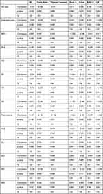
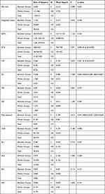
Corneal biomechanical parameters of the different myopia groups are shown in Table 1. There were statistically significant differences between the groups in the DA ratio (p = 0.033), SP-A (p=0.009), CBI (p=0.041), SSI (P=0.000), and Peak distance (p = 0.032). Bonferroni multiple comparisons between pairs are presented in Table 2. In correlation with different Corvis ST biomechanical variables, SE was found to be correlated with the DA ratio, SP-A, and SSI, as shown in Table 3 and Figure 1, while In the multiple regression analysis, SE was found to be independently correlated with SSI and peak distance, while the grade of myopia was found to be independently correlated with SP-A, as shown in Table 4.
 |
Table 1 Descriptive Analysis of Different Biomechanical Parameters in Each Myopia Group |
 |
Table 3 Correlation Between Different Biomechanical Variables, SE and Corneal Tomographic Variables |
 |
Table 4 Multivariate Regression Analysis Model Between Corneal Biomechanical Variables and SE, Grade of Myopia, IOP, CCT and Age |
Discussion
The management of myopia using refractive surgery is widely used; therefore, evaluation of corneal biomechanics has been used for the diagnosis and prediction of postoperative complications.12 A better understanding of corneal biomechanical characteristics in different grades of myopia might help improve the selection of suitable candidates for refractive procedures and guide the understanding of the management of biomechanic-modulating treatments.13,14 Many studies15,16–27 have been conducted to detect the associations between corneal biomechanics measurements using Corvis ST in China, India, and Iran with few studies incorporating extreme myopia cases and few have addressed the value of SSI. The current study is the first to describe corneal biomechanical properties in different grades of myopia in our country and found that SSI and Peak distance were independently correlated with refractive error spherical equivalent, and SP-A was independently correlated with the grade of myopia.
In the current study, IOP and CCT were correlated with most corneal biomechanical variables, as shown in Table 4, which is consistent with other studies.17,28,29 This explains the importance of the newly introduced SSI in the Corvis ST software. Because it assesses material stiffness, the invention of SSI solves this problem.30 In the analysis of the SSI parameter, Eliasy et al31 employed a numerical model that not only included a large range of changes in IOP, CCT, shape, and material parameters but also somewhat extended beyond the scope of clinical research. As a new measure independent of IOP and corneal geometry, SSI can detect high-risk or vulnerable individuals with ectasia after refractive surgery, alert clinicians to the potential hazards caused by decreased corneal biomechanical qualities, and aid in surgical planning. In the current study, we found that SE was independently correlated with SSI, and we found a statistically significant difference in SSI between the mild, moderate myopia, and extreme myopia groups, consistent with the findings of Han et al,18 who found that SSI values were lower in patients with higher grades of myopia; however, in their study, they included only two groups: the low myopia group with SE ≥ 3.00D and the high myopia group with SE > −6.00D, which is also consistent with other studies that involved extreme myopia patients among the Chinese population.23,27,
Peak distance (PD) is defined as the distance between two corneal peaks at the highest concavity.17 In our study, we found a statistically significant difference between the moderate, severe, and extreme myopia groups and found that SE was independently correlated with PD. These results are consistent with those of other studies,18,20,22 also Lu et al21 found a statistically significant correlation between axial length and PD in children; however, other studies testing the correlation between SE and PD found no significant results.27,28
The stiffness parameter at applanation 1 (SP-A1) represents corneal stiffness as derived from the resultant pressure divided by the deflection amplitude at applanation 1(A1).17 The more stiff the cornea, the more the value of SP-A1, in our study, we found that myopia grade was independently correlated with SP-A1 (coefficient p=−0.271, p=0.034), which is consistent with the results of other studies that analyzed SP-A1,27 while other studies did not show a significant difference between different myopic grades.17,2021 This finding could be explained by the different categorization of patients, as some studies categorized patients into only two groups, low to moderate myopia and high myopia groups, using the SE of −5D as a cut-off between both groups. In our study, we categorized patients into four groups to fully describe each. Another point of difference is the different ethnicities that could play a role in the differences in corneal biomechanics.
The deflection amplitude ratio (DA ratio) represents the resistance of the cornea to deformation; the greater the DA ratio, the lower is the resistance of the cornea to deformation. In our study, we found a higher DA ratio in patients with high SE than in those with lower ones, in the multivariate analysis, these results were insignificant. Our results were consistent with those of Kenia et al,17 who found comparable values of DA ratio in low to moderate myopia (4.48 ± 0.38), patients, high myopes (4.51 ± 0.42), and normal eyes (4.30 ± 0.50).32
The corneal biomechanical index (CBI) is a sensitive parameter for prediction of keratoconus and ectasia cases.30 The more biomechanically weaker the cornea, the closer the CBI value is to 1.32 In our study, we found only a statistically significant difference between the mild and moderate myopia groups, and CBI was correlated with SE; however, there was no correlation with SE or myopia grade in multivariate analysis. These results are consistent with those of other studies,17,20,2721 which did not find a significant correlation between SE and CBI. Therefore, CBI is affected more in cases of ectasia, regardless of refractive status.
In the current study, we tested the correlation between corneal tomographic parameters, namely, the Pachy apex, thinnest location, Kmax, mean K, and corneal volume (CV), as shown in Table 3 and found a significant correlation between these parameters and almost all parameters. These findings support the hypothesis that shape could affect corneal biomechanical assessment,33,34 and open a window to further investigate the interplay between tomography and biomechanics.
The strengths of our study come from the fact that it is the first to be conducted in this area, the prospective study design involving four myopia subgroups with inclusion of extreme myopia cases, and the proper analysis of the latest software of the CORVIS-ST device (software version 1.6r2187) that allowed further analysis of stiffness parameters such as SP-A and SSI. However, this study has a few limitations. Unfortunately, the number of eyes in the extreme myopia group was lower than in the other groups. In addition, we included only adult participants to limit the effect of age as a confounding variable, so we recommend further studies on the child age group with a larger sample size and follow-up studies to detect the effect of time on the progression of myopia and corneal biomechanics.
Conclusion
SSI, Peak distance, SP-A, and DA ratio are somehow correlated with SE, and the grade of myopia. We found the higher the grade of myopia; the less resistance to strain and more deformability of the cornea. The study confirms the need to address the biomechanics of the cornea before taking any critical decision regarding refractive surgery.
Data Sharing Statement
The datasets generated during and/or analyzed during the current study are available from the corresponding author on reasonable request.
Ethical Statement
This study was approved by the Central Scientific Research Ethical Committee Board of the High Council of University Hospitals number NO-0332[V2] and adhered to the Helsinki Declaration. Written informed consent was obtained from all the patients.
Funding
It is a self-funding study.
Disclosure
No conflict of interest for any of the authors.
References
1. Holden BA, Fricke TR, Wilson DA. Global prevalence of myopia and high myopia and temporal trends from 2000 through 2050. Ophthalmology. 2016;123(5):
2. Pesudovs K, Garamendi E, Elliott DB. A quality of life comparison of people wearing spectacles or contact lenses or having undergone refractive surgery. J Refract Surg. 2006;22(1):
3. Shams N, Mobaraki H, Kamali M, Jafarzadehpour E. Comparison of quality of life between myopic patients with spectacles and contact lenses, and patients who have undergone refractive surgery. J Curr Ophthalmol. 2015;27(1–2):
4. Long Q, Wang JY, Xu D, Li Y. Comparison of corneal biomechanics in Sjögren’s syndrome and non‐Sjogren’s syndrome dry eyes by scheimpflug based device. Int J Ophthalmol. 2017;10(5):
5. Luce DA. Determining in vivo biomechanical properties of the cornea with an ocular response analyzer. J Cataract Refract Surg. 2005;31(1):
6. Zhang J, Zheng L, Zhao X, Xu Y, Chen S. Corneal biomechanics after small‐incision lenticule extraction versus Q‐value‐guided femtosecond laser‐assisted in situ keratomileusis. J Curr Ophthalmol. 2016;28:
7. Lazreg S, Mesplié N, Praud D, et al. Comparison of corneal thickness and biomechanical properties between North African and French patients. J Cataract Refract Surg. 2013;39(3):
8. Vinciguerra R, Herber R, Wang Y. Corneal biomechanics differences between Chinese and Caucasian healthy subjects. Front Med. 2022;9:834663. doi:10.3389/fmed.2022.834663
9. Hilmi MR, Musa NH, Khairidzan MK, et al. Changes In Apical Corneal Curvature In Unilateral Primary Pterygium And Normal Adults Using Simulated-K And Corneal Irregularity Measurement. Int J Allied Health Sci. 2019;3(2):588–594.
10. Che Arif FA, Hilmi MR, Kamal MK, et al. Comparison of Immediate Effects on Usage of Dual Polymer Artificial Tears on Changes in Tear Film Characteristics. Malaysian J Med Health Sci. 2021;17(3):252–258.
11. Tang SM, Ma L, Lu SY, et al. Association of the PAX6 gene with extreme myopia rather than lower grade myopias. Br J Ophthalmol. 2018;102(4):570–574. doi:10.1136/bjophthalmol-2017-311327
12. Sedaghat MR, Momeni-Moghaddam H, Heravian J, et al. Detection ability of corneal biomechanical parameters for early diagnosis of ectasia. Eye (Lond)).2022;37:1665–1672
13. Flockerzi E, Xanthopoulou K, Daas L, et al. Evaluation of Dynamic Corneal Response Parameters and the Biomechanical E-Staging After Accelerated Corneal Cross-Linking in Keratoconus. Asia Pac J Ophthalmol (Phila). 2022;11(6):514–520. doi:10.1097/APO.0000000000000580
14. Xanthopoulou K, Seitz B, Belin MW, et al. Reliability analysis of successive Corvis ST(R) measurements in keratoconus 2 years after accelerated corneal crosslinking compared to untreated keratoconus corneas. Graefes Arch Clin Exp Ophthalmol. 2022;261(4):1055–1061. doi:10.1007/s00417-022-05881-6
15. Lee R, Chang RT, Wong IYH, et al. Assessment of corneal biomechanical parameters in myopes and emmetropes using the Corvis ST. Clin Exp Optom. 2016;99(2):157–162. doi:10.1111/cxo.12341
16. He M, Wang W, Ding H, et al. Corneal biomechanical properties in high myopia measured by dynamic scheimpflug imaging technology. Optom Vis Sci. 2017;94(12):
17. Kenia VP, Kenia RV, Pirdankar OH. Association between corneal biomechanical parameters and myopic refractive errors in young Indian individuals. Taiwan J Ophthalmol. 2020;10(1):45–53. doi:10.4103/tjo.tjo_15_19
18. Han F, Li M, Wei P, et al. Effect of biomechanical properties on myopia: a study of new corneal biomechanical parameters. BMC Ophthalmol. 2020;20(1):459. doi:10.1186/s12886-020-01729-x
19. Chu Z, Ren Q, Chen M, et al. The relationship between axial length/corneal radius of curvature ratio and stress-strain index in myopic eyeballs: using Corvis ST tonometry. Front Bioeng Biotechnol. 2022;10:939129. doi:10.3389/fbioe.2022.939129
20. Gao R, Ren Y, Li S, et al. Assessment of corneal biomechanics in anisometropia using Scheimpflug technology. Front Bioeng Biotechnol. 2022;10:994353. doi:10.3389/fbioe.2022.994353
21. Lu LL, Hu XJ, Yang Y, et al. Correlation of myopia onset and progression with corneal biomechanical parameters in children. World J Clin Cases. 2022;10:1548–1556. doi:10.12998/wjcc.v10.i5.1548
22. Sedaghat MR, Momeni-Moghaddam H, Azimi A, et al. Corneal biomechanical properties in varying severities of myopia. Front Bioeng Biotechnol. 2020;8:595330. doi:10.3389/fbioe.2020.595330
23. Liu Y, Pang C, Ming S, et al. Effect of myopia and astigmatism deepening on the corneal biomechanical parameter stress strain index in individuals of Chinese ethnicity. Front Bioeng Biotechnol. 2022;10:1018653. doi:10.3389/fbioe.2022.1018653
24. Liu G, Rong H, Zhang P, et al. The effect of axial length elongation on corneal biomechanical property. Front Bioeng Biotechnol. 2021;9:777239. doi:10.3389/fbioe.2021.777239
25. Wang J, Li Y, Jin Y, et al. Corneal biomechanical properties in myopic eyes measured by a Dynamic Scheimpflug Analyzer. J Ophthalmol. 2015;2015:161869. doi:10.1155/2015/161869
26. Long W, Zhao Y, Hu Y, et al. Characteristics of corneal biomechanics in Chinese preschool children with different refractive status. Cornea. 2019;38(11):1395–1399. doi:10.1097/ICO.0000000000001971
27. Li DL, Liu MX, Yin ZJ, et al. Refractive associations with corneal biomechanical properties among young adults: a population-based Corvis ST study. Graefes Arch Clin Exp Ophthalmol. 2023. doi:10.1007/s00417-023-06164-4
28. He M, Ding H, He H, et al. Corneal biomechanical properties in healthy children measured by corneal visualization scheimpflug technology. BMC Ophthalmol. 2017;17(1):70. doi:10.1186/s12886-017-0463-x
29. Wang W, He M, He H, Zhang C, Jin H, Zhong X. Corneal biomechanical metrics of healthy Chinese adults using Corvis ST. Cont Lens Anterior Eye. 2017;40(2):97–103. doi:10.1016/j.clae.2016.12.003
30. Vinciguerra R, Ambrosio RJ, Elsheikh A, et al. Detection of Keratoconus with a new biomechanical index. J Refract Surg. 2016;32(12):803–810. doi:10.3928/1081597X-20160629-01
31. Eliasy A, Chen KJ, Vinciguerra R, et al. Ex-vivo experimental validation of biomechanically-corrected intraocular pressure measurements on human eyes using the CorVis ST. Exp Eye Res. 2018;175:98–102. doi:10.1016/j.exer.2018.06.013
32. Ambrósio R, Lopes BT, Faria-Correia F, et al. Integration of scheimpflug based corneal tomography and biomechanical assessments for enhancing ectasia detection. J Refract Surg. 2017;33(7):434–443. doi:10.3928/1081597X-20170426-02
33. Zhang L, Wang Y, Xie L, et al. The Relationship between Corneal Biomechanics and Corneal Shape in Normal Myopic Eyes. J Clin Exp Ophthalmol. 2013;4(03):278. doi:10.4172/2155-9570.1000278
34. Wilson A, Marshall J. A review of corneal biomechanics: mechanisms for measurement and the implications for refractive surgery. Indian J Ophthalmol. 2020;68(12):2679–2690. doi:10.4103/ijo.IJO_2146_20
 © 2024 The Author(s). This work is published and licensed by Dove Medical Press Limited. The
full terms of this license are available at https://www.dovepress.com/terms.php
and incorporate the Creative Commons Attribution
- Non Commercial (unported, v3.0) License.
By accessing the work you hereby accept the Terms. Non-commercial uses of the work are permitted
without any further permission from Dove Medical Press Limited, provided the work is properly
attributed. For permission for commercial use of this work, please see paragraphs 4.2 and 5 of our Terms.
Recommended articles
Scheimpflug-Based Corneal Biomechanical Analysis As A Predictor of Glaucoma in Eyes With High Myopia
Baptista PML, Ferreira AS, Silva NP, Figueiredo ARM, Sampaio IC, Reis RVF, Ambrósio R Jr, Menéres PMAM, Beirão JNM, Menéres MJFS
Clinical Ophthalmology 2024, 18:545-563
Published Date: 22 February 2024


